Accéder au contenu
Merck
Toutes les photos(1)

Principaux documents

A5968

Sigma-Aldrich

Anti-AP-1 antibody produced in rabbit

affinity isolated antibody, buffered aqueous solution

Synonyme(s) :

Ap-1 Antibody, Ap1 Antibody, Ap1 Antibody - Anti-AP-1 antibody produced in rabbit, Anti-c-Jun

Se connecterpour consulter vos tarifs contractuels et ceux de votre entreprise/organisme


About This Item

Numéro MDL:
Code UNSPSC :
12352203
Nomenclature NACRES :
NA.41
Conjugué:
unconjugated
application:
ARR
IHC (p)
WB
Clone:
polyclonal
Espèces réactives:
human
citations:
18
Technique(s):
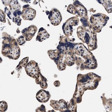
immunohistochemistry (formalin-fixed, paraffin-embedded sections): 1:200 using formalin-fixed, paraffin-embedded sections of human colon carcinoma
microarray: suitable
western blot: 1:200 using HeLa nuclear extract

Source biologique

rabbit

Niveau de qualité

Conjugué

unconjugated

Forme d'anticorps

affinity isolated antibody

Type de produit anticorps

primary antibodies

Clone

polyclonal

Forme

buffered aqueous solution

Poids mol.

antigen 39 kDa

Espèces réactives

human

Technique(s)

immunohistochemistry (formalin-fixed, paraffin-embedded sections): 1:200 using formalin-fixed, paraffin-embedded sections of human colon carcinoma
microarray: suitable
western blot: 1:200 using HeLa nuclear extract

Numéro d'accès UniProt

Conditions d'expédition

dry ice

Température de stockage

−20°C

Modification post-traductionnelle de la cible

unmodified

Informations sur le gène

human ... JUN(3725)

Description générale

Activation Protein transcription factor 1 (AP-1) is a leucine zipper transcription factor that recognizes a specific palindromic sequence present in several promoters and enhancers. AP-1 can be induced by several growth and apoptosis-related signaling pathways. Jun proteins are also involved in metastatic, inflammatory and stress responses.
Anti-AP-1/c-Jun recognizes an epitope located on the c-Jun DNA binding domain. This epitope is highly conserved in c-Jun, Jun B and Jun D proteins of chicken, mouse, rat, and human. By immunoblotting, the antibody reacts specifically with AP-1/c-Jun (a single band or occasionally a doublet at 39kDa region). Additional bands of lower molecular weight may be observed. Staining of AP-1/c-Jun band(s) is inhibited by the AP-1/c-Jun immunizing peptide (amino acid residues 246-263).
Activation Protein transcription factor 1 (AP-1) is a regulatory leucine zipper transcription factor. AP-1 is composed of either a homodimer of two Jun protein molecules or a heterodimer of one Jun and one Fos (Proto-oncogenes) molecules.

Immunogène

synthetic peptide corresponding to amino acids 246-263 of human c-Jun with N-terminally added lysine, conjugated to KLH.

Application

Anti-AP-1 antibody has been used in
  • immunohistochemistry
  • immunochemistry
  • western blotting

Immunofluorescence of Hela cells was performed using monoclonal anti-AP1 (clone 100/3) as the primary antibody.

Actions biochimiques/physiologiques

Activation Protein transcription factor 1 (AP-1) recognizes a specific sequence (TPA response element, a palindromic TRE sequence TGA(C/G) TCA or related sequences) present in many promoter and enhancer gene regions. Regulation of AP-1 is achieved by changes in the expression of Jun and Fos proteins in the dimer, by phosphorylation at both Ser63 and Ser73 by c-Jun N-terminal kinases (JNK) and by interaction with a variety of transcriptional coactivators like C-Jun activation domain-binding protein-1 (JAB1). AP-1 is induced by a variety of signals including those eliciting proliferation, differentiation and apoptosis.

Forme physique

Solution in 0.01 M phosphate buffered saline, pH 7.4, containing 1% bovine serum albumin and 15 mM sodium azide.

Clause de non-responsabilité

Unless otherwise stated in our catalog or other company documentation accompanying the product(s), our products are intended for research use only and are not to be used for any other purpose, which includes but is not limited to, unauthorized commercial uses, in vitro diagnostic uses, ex vivo or in vivo therapeutic uses or any type of consumption or application to humans or animals.

Vous ne trouvez pas le bon produit ?  

Essayez notre Outil de sélection de produits.

Code de la classe de stockage

10 - Combustible liquids

Classe de danger pour l'eau (WGK)

WGK 3

Point d'éclair (°F)

Not applicable

Point d'éclair (°C)

Not applicable


Faites votre choix parmi les versions les plus récentes :

Certificats d'analyse (COA)

Lot/Batch Number

Vous ne trouvez pas la bonne version ?

Si vous avez besoin d'une version particulière, vous pouvez rechercher un certificat spécifique par le numéro de lot.

Déjà en possession de ce produit ?

Retrouvez la documentation relative aux produits que vous avez récemment achetés dans la Bibliothèque de documents.

Consulter la Bibliothèque de documents

Twelve hours of heat stress induces inflammatory signaling in porcine skeletal muscle
Ganesan S, et al.
The American Journal of Physiology, 310(11), R1288-R1296 (2016)
Ui Joung Youn et al.
Biological & pharmaceutical bulletin, 34(6), 890-893 (2011-06-02)
Tumor necrosis factor α (TNF-α), which is a primary cytokine responsible for inflammatory responses in skin, induces the synthesis of matrix metalloproteinase-9 (MMP-9), which causes skin aging. The protective effects of 3-deoxysappanchalcone against TNF-α-induced damage was investigated using human skin
Swathy Krishna et al.
American journal of physiology. Regulatory, integrative and comparative physiology, 325(6), R692-R711 (2023-10-09)
Duchenne muscular dystrophy (DMD), a progressive muscle disease caused by the absence of functional dystrophin protein, is associated with multiple cellular, physiological, and metabolic dysfunctions. As an added complication to the primary insult, obesity/insulin resistance (O/IR) is frequently reported in
Vasilios P Koulouras et al.
International journal of clinical and experimental medicine, 4(1), 53-66 (2011-03-12)
Recent animal studies have demonstrated that pre-treatment with inhaled carbon monoxide (iCO) exert anti-inflammatory effects in various septic models. In all these models, there is no information whether iCO might act therapeutically after the onset of septic damage. The objective
Samuel Jang et al.
Cancer medicine, 6(9), 2142-2152 (2017-08-05)
Notch signaling is minimally active in neuroendocrine (NE) cancer cells. While histone deacetylase inhibitors (HDACi) suppress NE cancer growth by inducing Notch, the molecular mechanism underlying this interplay has not yet been defined. NE cancer cell lines BON, H727, and

Notre équipe de scientifiques dispose d'une expérience dans tous les secteurs de la recherche, notamment en sciences de la vie, science des matériaux, synthèse chimique, chromatographie, analyse et dans de nombreux autres domaines..

Contacter notre Service technique