Skip to Content
Merck
All Photos(7)

Key Documents

L8918

Sigma-Aldrich

Anti-LC3 antibody produced in rabbit

~1 mg/mL, affinity isolated antibody, buffered aqueous solution

Synonym(s):

Anti-Lc3, LC3 Antibody - Anti-LC3 antibody produced in rabbit, Lc3 Antibody, Anti-MAP1 light chain 3-like protein 1, Anti-MAP1A/1B light chain 3A, Anti-MAP1A/1BLC3, Anti-MAP1A/MAP1B LC3A, Anti-MAP1ALC3, Anti-MAP1BLC3, Anti-MAP1LC3A, Anti-MAP1LC3B, Anti-Microtubule-associated protein 1 light chain 3 alpha, Anti-Microtubule-associated protein 1 light chain 3 beta, Anti-Microtubule-associated proteins 1A/1B light chain 3

Sign Into View Organizational & Contract Pricing


About This Item

UNSPSC Code:
12352203
NACRES:
NA.41

biological source

rabbit

Quality Level

conjugate

unconjugated

antibody form

affinity isolated antibody

antibody product type

primary antibodies

clone

polyclonal

form

buffered aqueous solution

mol wt

antigen 16-18 kDa

species reactivity

mouse, human, rat

packaging

antibody small pack of 25 μL

concentration

~1 mg/mL

technique(s)

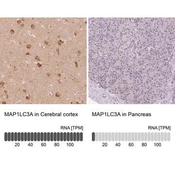
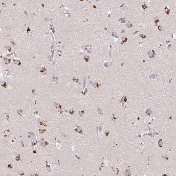
immunohistochemistry: suitable
immunoprecipitation (IP): 5-10 μg using extracts of human U87 cells
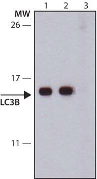
western blot: 1-2 μg/mL using whole extracts of rat and mouse brain

UniProt accession no.

shipped in

dry ice

storage temp.

−20°C

target post-translational modification

unmodified

General description

The gene LC3, also referred to as MAP1LC3A (microtubule associated protein 1 light chain 3 α), is a homologue of Apg8p necessary for autophagy in yeast. The cytosolic form upon processing gets localized to the autophagosome membranes. The gene is mapped to human chromosome 20q11.22. LC3 exists in cells as cytoplasmic LC3-I (18kDa) and LC3-II (16kDa).

Immunogen

Synthetic peptide corresponding to amino acids of human LC3A isoform a, conjugated to KLH via C-terminal cysteine residue. The corresponding sequence is identical in rat and mouse.

Application

Anti-LC3 antibody produced in rabbit has been used in:
  • western blotting
  • immunofluorescence
  • immunohistochemistry
  • filter retardation assay
  • immunocytochemistry
Applications in which this antibody has been used successfully, and the associated peer-reviewed papers, are given below.
Immunofluorescence (3 papers)
Immunohistochemistry (1 paper)
Western Blotting (21 paper)

Biochem/physiol Actions

The gene MAP1LC3A (microtubule associated protein 1 light chain 3 α) encodes a protein that serves as a marker of autophagosomes. It is found to be expressed on surviving oligodendrocytes in Nasu-Hakola disease brains and may be involved in the induction of oligodendrogliopathy.

Physical form

Solution in 0.01 M phosphate buffered saline, pH 7.4, containing 15 mM sodium azide.

Storage and Stability

For continuous use, store at 2-8 °C for up to one month. For extended storage freeze in working aliquots. Repeated freezing and thawing is not recommended. If slight turbidity occurs upon prolonged storage, clarify the solution by centrifugation before use. Working dilution samples should be discarded if not used within 12 hours.

Disclaimer

Unless otherwise stated in our catalog or other company documentation accompanying the product(s), our products are intended for research use only and are not to be used for any other purpose, which includes but is not limited to, unauthorized commercial uses, in vitro diagnostic uses, ex vivo or in vivo therapeutic uses or any type of consumption or application to humans or animals.

Not finding the right product?  

Try our Product Selector Tool.

Storage Class Code

10 - Combustible liquids

WGK

WGK 3

Personal Protective Equipment

dust mask type N95 (US), Eyeshields, Gloves

Certificates of Analysis (COA)

Search for Certificates of Analysis (COA) by entering the products Lot/Batch Number. Lot and Batch Numbers can be found on a product’s label following the words ‘Lot’ or ‘Batch’.

Already Own This Product?

Find documentation for the products that you have recently purchased in the Document Library.

Visit the Document Library

LC3, an autophagosome marker, is expressed on oligodendrocytes in Nasu-Hakola disease brains.
Satoh J
Orphanet Journal of Rare Diseases, 9 (2014)
Peng Yin et al.
PloS one, 10(12), e0143922-e0143922 (2015-12-05)
We investigated the mechanisms underlying damage to rat small intestine in heat- and shake-induced stress. Eighteen Sprague-Dawley rats were randomly divided into a control group and a 3-day stressed group treated 2 h daily for 3 days on a rotary
TRIAD1 Is a Novel Transcriptional Target of p53 and Regulates Nutlin-3a-Induced Cell Death.
Lee J
Journal of Cellular Biochemistry (2016)
Benoît Renvoisé et al.
Annals of clinical and translational neurology, 1(6), 379-389 (2014-07-08)
Hereditary spastic paraplegias (HSPs) are among the most genetically diverse inherited neurological disorders, with over 70 disease loci identified (SPG1-71) to date. SPG15 and SPG11 are clinically similar, autosomal recessive disorders characterized by progressive spastic paraplegia along with thin corpus
Pengfei Jiang et al.
Journal of translational medicine, 12, 13-13 (2014-01-18)
Glioblastoma (GBM) is a therapeutic challenge, associated with high mortality. More effective GBM therapeutic options are urgently needed. Hence, we screened a large multi-class drug panel comprising the NIH clinical collection (NCC) that includes 446 FDA-approved drugs, with the goal

Articles

High titer lentiviral particles for LC3 variants used for live cell analysis of cellular autophagy.

Our team of scientists has experience in all areas of research including Life Science, Material Science, Chemical Synthesis, Chromatography, Analytical and many others.

Contact Technical Service