Skip to Content
Merck
All Photos(3)

Key Documents

SCC173

Sigma-Aldrich

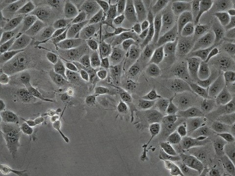
MCA205 Mouse Fibrosarcoma Cell Line

Mouse

Synonym(s):

MCA-205, MCA 205

Sign Into View Organizational & Contract Pricing


About This Item

UNSPSC Code:
41106514
eCl@ss:
32011203
NACRES:
NA.81

product name

MCA205 Mouse Fibrosarcoma Cell Line, MCA205 mouse fibrosarcoma cell line is an excellent model for studying immune response to tumor cells and the development of targeted cancer immunotherapies.

biological source

mouse

technique(s)

cell culture | mammalian: suitable

shipped in

ambient

General description

Cancer immunotherapy is a rapidly expanding field that has produced significant advances in cancer treatment. Understanding the interactions of tumor cells with the immune system is crucial to the development of effective immunotherapeutic approaches. Fibrosarcoma is a rare yet aggressive soft-tissue sarcoma characterized by frequent tumor recurrences and a high degree of resistance to radio- and chemotherapy .
MCA-205 is a weakly immunogenic fibrosarcoma-derived cell line that has been used in many studies to induce tumor-reactive T lymphocytes, the key aspect of adoptive immunotherapy . MCA-205 cells are widely utilized as stimulators for inducing cytokine expression . The MCA-205 mouse fibrosarcoma cell line is an excellent model for studying the immune responses to tumor cells and for supporting the development of targeted cancer immunotherapies.

Source:
Non-GMO. MCA-205 was derived from 3-methylcholanthrene-induced fibrosarcoma in C57BL/6 mice. Tumors were maintained in vivo by serial subcutaneous transplantation in syngeneic mice and single-cell suspensions were prepared from solid tumors by enzymatic digestion . From these cells the MCA-205 cell line was established and maintained in vitro.

Cell Line Description

Cancer Cells

Application

MCA205 mouse fibrosarcoma cell line is an excellent model for studying immune response to tumor cells and the development of targeted cancer immunotherapies.
Research Category
Cancer

Oncology
This product is sold solely for research use per the terms of the “Restricted Use Agreement” which govern its use as detailed in the product Data Sheet or Certificate of Analysis. For information regarding any other use, please contact licensing@emdmillipore.com.

Quality

• Each vial contains ≥ 1X10⁶ viable cells.
• Cells are tested negative for infectious diseases by a Mouse Essential CLEAR panel by Charles River Animal Diagnostic Services.
• Cells are verified to be of mouse origin and negative for inter-species contamination from rat, chinese hamster, Golden Syrian hamster, human and non-human primate (NHP) as assessed by a Contamination CLEAR panel by Charles River Animal Diagnostic Services.
• Cells are negative for mycoplasma contamination

Storage and Stability

Store in liquid nitrogen. The cells can be cultured for at least 10 passages after initial thawing without significantly affecting the cell marker expression and functionality.

Disclaimer

Unless otherwise stated in our catalog or other company documentation accompanying the product(s), our products are intended for research use only and are not to be used for any other purpose, which includes but is not limited to, unauthorized commercial uses, in vitro diagnostic uses, ex vivo or in vivo therapeutic uses or any type of consumption or application to humans or animals.

Storage Class Code

12 - Non Combustible Liquids

WGK

WGK 2

Flash Point(F)

Not applicable

Flash Point(C)

Not applicable


Certificates of Analysis (COA)

Search for Certificates of Analysis (COA) by entering the products Lot/Batch Number. Lot and Batch Numbers can be found on a product’s label following the words ‘Lot’ or ‘Batch’.

Already Own This Product?

Find documentation for the products that you have recently purchased in the Document Library.

Visit the Document Library

H Kagamu et al.
Cancer research, 56(19), 4338-4342 (1996-10-01)
The ability to generate a large number of tumor-reactive T lymphocytes is the most critical requirement for adoptive immunotherapy. Our laboratory has previously demonstrated that cells from tumor-draining lymph nodes (LNs) are an excellent source of tumor-reactive T lymphocytes. After
T Chou et al.
Journal of immunology (Baltimore, Md. : 1950), 140(7), 2453-2461 (1988-04-01)
We have previously established an in vitro sensitization (IVS) procedure with which lymphocytes from tumor-bearing mice could be expanded and sensitized to acquire antitumor reactivity capable of mediating the regression of established pulmonary metastases from the weakly immunogenic MCA 105
Klaire Yixin Fjæstad et al.
Oncogene, 41(9), 1364-1375 (2022-01-13)
The development of immune checkpoint inhibitors (ICI) marks an important breakthrough of cancer therapies in the past years. However, only a limited fraction of patients benefit from such treatments, prompting the search for immune modulating agents that can improve the
R J Barth et al.
Journal of immunology (Baltimore, Md. : 1950), 144(4), 1531-1537 (1990-02-15)
Tumor infiltrating lymphocytes (TIL) were cultured from multiple methylcholanthrene-induced sarcomas under four different conditions: either low-dose (10 U/ml) or high-dose (1000 U/ml) rIL-2 was used with Ag stimulation by irradiated autologous tumor and splenocytes starting either at day 1 or
Myrofora Panagi et al.
Nature communications, 13(1), 7165-7165 (2022-11-24)
Nano-immunotherapy improves breast cancer outcomes but not all patients respond and none are cured. To improve efficacy, research focuses on drugs that reprogram cancer-associated fibroblasts (CAFs) to improve therapeutic delivery and immunostimulation. These drugs, however, have a narrow therapeutic window

Our team of scientists has experience in all areas of research including Life Science, Material Science, Chemical Synthesis, Chromatography, Analytical and many others.

Contact Technical Service