Skip to Content
Merck
All Photos(1)

Key Documents

P7607

Sigma-Aldrich

Monoclonal Anti-Protein Phosphatase 1α antibody produced in mouse

clone PPI-377, ascites fluid

Synonym(s):

Anti-PP1α

Sign Into View Organizational & Contract Pricing


About This Item

MDL number:
UNSPSC Code:
12352203
NACRES:
NA.44

biological source

mouse

Quality Level

conjugate

unconjugated

antibody form

ascites fluid

antibody product type

primary antibodies

clone

PPI-377, monoclonal

mol wt

antigen 37.5 kDa

contains

15 mM sodium azide

species reactivity

rabbit, rat, bovine, human, mouse, monkey

technique(s)

immunocytochemistry: suitable
microarray: suitable
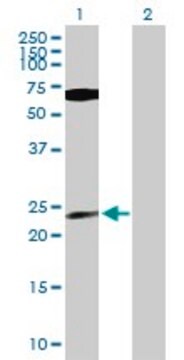
western blot: 1:500 using mouse fibroblasts cell extract

isotype

IgG2b

UniProt accession no.

shipped in

dry ice

storage temp.

−20°C

target post-translational modification

unmodified

Gene Information

human ... PPP1CA(5499)
mouse ... Ppp1ca(19045)
rat ... Ppp1ca(24668)

General description

Among the post-translational modifications, phosphorylation is a vital regulatory mechanism of key proteins involved in specific pathways. Reverse phosphorylation has become recognized as the key process of regulation of gene expression, cellular proliferation, differentiation in Eukaryotes. Protein phosphatases, like kinases, are a class of enzymes that regulate protein phosphorylation. The serine/threonine phosphatases have been classified into four groups which include PP1, PP2A, PP2B (also termed calcineurin) and PP2C on the basis of differences in their biochemical properties. PP1 catalyzes a wide range of protein dephosphorylation reactions in a tightly regulated manner and is expressed abundantly in the brain. PP1 has broad functions covering glycogen metabolism, protein synthesis, cell cycle and growth and muscle contractility. PP1 forms exclusive complexes with >50 regulatory subunits that allows for restricted subcellular location and thereby distinct cellular functions. There are 3 isoforms of PP1 (α, β and γ1) with 90% homology. PP1α is expressed in brain, specifically in cerebellum, prefrontal cortex and synapses
Monoclonal Anti-Protein Phosphatase 1α specifically recognizes an epitope within the catalytic subunit of PP1α isoform (37.5 kDa).

Specificity

The antibody reacts with protein phosphatase 1α and recognizes an epitope within its catalytic subunit.

Immunogen

recombinant rabbit protein phosphatase 1α (PP1α) catalytic subunit

Application

Anti-Protein Phosphatase 1α is suitable for immunoblotting at a working dilution of 1:500 using mouse fibroblasts cell extract. A dilution of 1:200 has been used for immunoblotting in myocardial protein extracts of Göttinger minipigs. For immunoprecipitation, 1 μg antibody/20 μg of pig protein extracts was suitable. The antibody is also suitable for immunocytochemistry and protein microarray.
Applications in which this antibody has been used successfully, and the associated peer-reviewed papers, are given below.
Western Blotting (1 paper)

Disclaimer

Unless otherwise stated in our catalog or other company documentation accompanying the product(s), our products are intended for research use only and are not to be used for any other purpose, which includes but is not limited to, unauthorized commercial uses, in vitro diagnostic uses, ex vivo or in vivo therapeutic uses or any type of consumption or application to humans or animals.

Not finding the right product?  

Try our Product Selector Tool.

Storage Class Code

10 - Combustible liquids

WGK

nwg

Flash Point(F)

Not applicable

Flash Point(C)

Not applicable


Choose from one of the most recent versions:

Certificates of Analysis (COA)

Lot/Batch Number

Don't see the Right Version?

If you require a particular version, you can look up a specific certificate by the Lot or Batch number.

Already Own This Product?

Find documentation for the products that you have recently purchased in the Document Library.

Visit the Document Library

Jill R Bordelon et al.
Cerebral cortex (New York, N.Y. : 1991), 15(12), 1928-1937 (2005-03-11)
Prefrontal cortical functioning depends on D1 family receptors and their complex signal transduction cascade, including protein phosphatase-1 (PP1). Three PP1 isoforms are prominent in the brain: PP1alpha, PP1beta and PP1gamma1. PP1 localization by a variety of scaffolding proteins is critical
Differential cellular and subcellular localization of protein phosphatase 1 isoforms in brain
Strack S et al
The Journal of Comparative Neurology, 25, 373-384 (1999)
A Fischer et al.
Toxicology and applied pharmacology, 245(1), 9-20 (2010-02-23)
Cellular uptake of microcystins (MCs), a family of cyclic cyanobacterial heptapeptide toxins, occurs via specific organic anion transporting polypeptides (OATPs), where MCs inhibit serine/threonine-specific protein phosphatase (PP). Despite comparable PP-inhibitory capacity, MCs differ greatly in their acute toxicity, thus raising
Andreas Totzeck et al.
American journal of physiology. Heart and circulatory physiology, 295(5), H2106-H2112 (2008-10-07)
Cardiac connexin 43 (Cx43) is involved in infarct propagation, and the uncoupling of Cx43-formed channels reduces infarct size. Cx43-formed channels open upon Cx43 dephosphorylation, and ischemic preconditioning (IP) prevents the ischemia-induced Cx43 dephosphorylation. In addition to the sarcolemma, Cx43 is
Mathieu Bollen et al.
Trends in biochemical sciences, 35(8), 450-458 (2010-04-20)
Protein Ser/Thr phosphatase-1 (PP1) catalyzes the majority of eukaryotic protein dephosphorylation reactions in a highly regulated and selective manner. Recent studies have identified an unusually diversified PP1 interactome with the properties of a regulatory toolkit. PP1-interacting proteins (PIPs) function as

Our team of scientists has experience in all areas of research including Life Science, Material Science, Chemical Synthesis, Chromatography, Analytical and many others.

Contact Technical Service