Accéder au contenu
Merck
Toutes les photos(7)

Principaux documents

HPA002643

Sigma-Aldrich

Anti-CASP3 antibody produced in rabbit

enhanced validation

Prestige Antibodies® Powered by Atlas Antibodies, affinity isolated antibody, buffered aqueous glycerol solution

Synonyme(s) :

Anti-Apopain, Anti-CASP-3, Anti-CPP-32, Anti-Caspase-3 precursor, Anti-Cysteine protease CPP32, Anti-SCA-1, Anti-SREBP cleavage activity 1, Anti-Yama protein

Se connecterpour consulter vos tarifs contractuels et ceux de votre entreprise/organisme


About This Item

Numéro MDL:
Code UNSPSC :
12352203
Numéro HPA (Human Protein Atlas):
Nomenclature NACRES :
NA.41

Source biologique

rabbit

Niveau de qualité

Conjugué

unconjugated

Forme d'anticorps

affinity isolated antibody

Type de produit anticorps

primary antibodies

Clone

polyclonal

Gamme de produits

Prestige Antibodies® Powered by Atlas Antibodies

Forme

buffered aqueous glycerol solution

Espèces réactives

human

Validation améliorée

orthogonal RNAseq
Learn more about Antibody Enhanced Validation

Technique(s)

immunoblotting: 0.04-0.4 μg/mL
immunofluorescence: 0.25-2 μg/mL
immunohistochemistry: 1:1000-1:2500

Séquence immunogène

HGSESMDSGISLDNSYKMDYPEMGLCIIINNKNFHKSTGMTSRSGTDVDAANLRETFRNLKYEVRNKNDLTREEIVELMRDVSKEDHSKRSSFVCVLLSHGEEGIIFGTNGPVDL

Numéro d'accès UniProt

Conditions d'expédition

wet ice

Température de stockage

−20°C

Modification post-traductionnelle de la cible

unmodified

Informations sur le gène

human ... CASP3(836)

Description générale

CASP3 (caspase 3), the allosteric regulator, is a member of the cysteine-aspartic acid protease (caspase) family which is involved in the inflammation and mammalian apoptosis. It consists of binding sites for small molecules and peptides.

Immunogène

Caspase-3 precursor recombinant protein epitope signature tag (PrEST)

Application

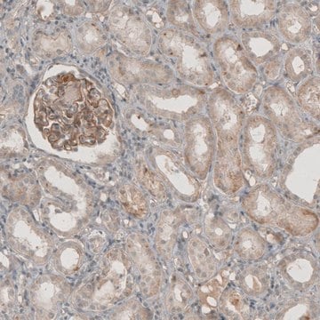
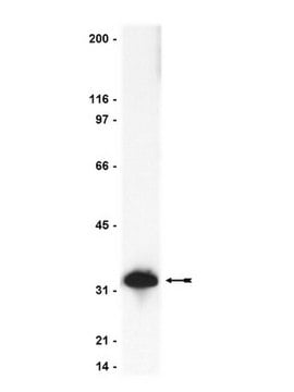
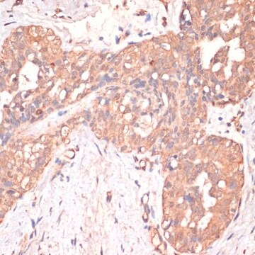
Anti-CASP3 antibody produced in rabbit, a Prestige Antibody, is developed and validated by the Human Protein Atlas (HPA) project . Each antibody is tested by immunohistochemistry against hundreds of normal and disease tissues. These images can be viewed on the Human Protein Atlas (HPA) site by clicking on the Image Gallery link. The antibodies are also tested using immunofluorescence and western blotting. To view these protocols and other useful information about Prestige Antibodies and the HPA, visit sigma.com/prestige.

Actions biochimiques/physiologiques

CASP3 (caspase 3) may have an impact on the melanoma tumor growth after the cytotoxic therapy. It plays a vital role in the proliferation of surrounding cells during executioner phase of apoptosis. Caspases are synthesized and localized as inactive zymogens. After a cascade of proteolytic processing they get activated. Upon activation, they cleave into two separate subunits which further dimerize to form the active enzyme. The activity of caspase-3 is induced by the accumulation of reactive oxygen species (ROS), mitochondrial dysfunction and reduction in adenosine triphosphate (ATP) levels. Alteration in the CASP3 gene is associated with neuronal death in Alzheimer′s disease.

Caractéristiques et avantages

Prestige Antibodies® are highly characterized and extensively validated antibodies with the added benefit of all available characterization data for each target being accessible via the Human Protein Atlas portal linked just below the product name at the top of this page. The uniqueness and low cross-reactivity of the Prestige Antibodies® to other proteins are due to a thorough selection of antigen regions, affinity purification, and stringent selection. Prestige antigen controls are available for every corresponding Prestige Antibody and can be found in the linkage section.

Every Prestige Antibody is tested in the following ways:
  • IHC tissue array of 44 normal human tissues and 20 of the most common cancer type tissues.
  • Protein array of 364 human recombinant protein fragments.

Liaison

Corresponding Antigen APREST86566

Forme physique

Solution in phosphate-buffered saline, pH 7.2, containing 40% glycerol and 0.02% sodium azide

Informations légales

Prestige Antibodies is a registered trademark of Merck KGaA, Darmstadt, Germany

Clause de non-responsabilité

Unless otherwise stated in our catalog or other company documentation accompanying the product(s), our products are intended for research use only and are not to be used for any other purpose, which includes but is not limited to, unauthorized commercial uses, in vitro diagnostic uses, ex vivo or in vivo therapeutic uses or any type of consumption or application to humans or animals.

Vous ne trouvez pas le bon produit ?  

Essayez notre Outil de sélection de produits.

Code de la classe de stockage

10 - Combustible liquids

Classe de danger pour l'eau (WGK)

WGK 1

Point d'éclair (°F)

Not applicable

Point d'éclair (°C)

Not applicable

Équipement de protection individuelle

Eyeshields, Gloves, multi-purpose combination respirator cartridge (US)


Certificats d'analyse (COA)

Recherchez un Certificats d'analyse (COA) en saisissant le numéro de lot du produit. Les numéros de lot figurent sur l'étiquette du produit après les mots "Lot" ou "Batch".

Déjà en possession de ce produit ?

Retrouvez la documentation relative aux produits que vous avez récemment achetés dans la Bibliothèque de documents.

Consulter la Bibliothèque de documents

Y Kitamura et al.
Brain research, 780(2), 260-269 (1998-03-21)
Recently, apoptosis has been implicated in the selective neuronal loss of Alzheimer's disease (AD). Apoptosis is regulated by the B cell leukemia-2 gene product (Bcl-2) family (Bcl-2, Bcl-x, Bax, Bak and Bad) and the caspase family (ICH-1 and CPP32), with
Maria Ana Contín et al.
Molecular vision, 19, 1614-1625 (2013-08-01)
Retinal degeneration caused by a defect in the phototransduction cascade leads to the apoptosis of photoreceptor cells, although the precise molecular mechanism is still unknown. In addition, constant low light exposure produces photoreceptor cell death through the activation of downstream
Connexin 43 and Its Hemichannels Mediate Hypoxia?Ischemia-Induced Cell Death in Neonatal Rats
Wang, J
Journal of Child Neurology, 1(1), 1-9 (2014)
Comparative pathology and immunohistochemistry of Newcastle disease in domestic chicken (Gallus-gallus domesticus) and Alabio duck (Anas platyrhynchos Borneo).
Etriwati, et al.
Open veterinary journal, 13, 433-442 (2023)
Alessandro Rava et al.
Journal of cellular physiology, 237(12), 4563-4579 (2022-11-03)
The loss of NPC1 or NPC2 function results in cholesterol and sphingolipid dyshomeostasis that impairs developmental trajectories, predisposing the postnatal brain to the appearance of pathological signs, including progressive and stereotyped Purkinje cell loss and microgliosis. Despite increasing evidence reporting

Notre équipe de scientifiques dispose d'une expérience dans tous les secteurs de la recherche, notamment en sciences de la vie, science des matériaux, synthèse chimique, chromatographie, analyse et dans de nombreux autres domaines..

Contacter notre Service technique