C-12217
Human Dermal Lymphatic Endothelial Cells (HDLEC)
Adult, 500,000 cryopreserved cells
Synonyme(s) :
HDLEC cells
Se connecterpour consulter vos tarifs contractuels et ceux de votre entreprise/organisme
About This Item
Code UNSPSC :
41106514
Nomenclature NACRES :
NA.81
Produits recommandés
Source biologique
human dermis
Conditionnement
pkg of 500,000 cells
Morphologie
( endothelial)
Technique(s)
cell culture | mammalian: suitable
Conditions d'expédition
dry ice
Température de stockage
−196°C
Description générale
Lot specific orders are not able to be placed through the web. Contact your local sales rep for more details.
Origine de la lignée cellulaire
Skin
Application
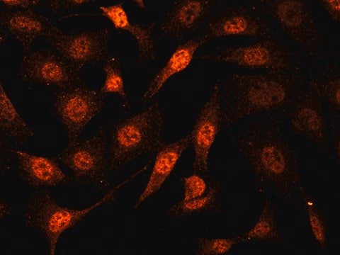
Primary Human Dermal Lymphatic Endothelial Cells (HDLEC) are a subpopulation of the Human Dermal Endothelial Cells. They are isolated from the dermis of adult skin (different locations) from a single donor and are provided in a cryopreserved format. (Human Dermal Blood Endothelial Cells (HDBEC) from the same donor are available on request.)The cells are routinely analyzed by flow cytometric analysis and immunofluorescent staining: >95% of the cells are CD31 positive and podoplanin positive. In addition, the cells stain positive for Prox-1.Lymphatic vessels transport excess fluids from tissues to the circulatory system and are a major component of the immune system. The transported fluid is nearly cell free. The vessels are highly permeable, because they lack a continuous basal membrane. Lymphatic endothelial cells are involved in pathological alterations of the lymphatic system. During tumor lymphangiogenesis, the lymphatic endothelial cells build new vessels that infiltrate tumors, attract tumor cells, and induce tumor cell metastasis.
Qualité
Rigid quality control tests are performed for each lot of Microvascular Endothelial Cells. They are tested for cell morphology and cell type specific markers, e.g. von Willebrand Factor (vWF), CD31 and Podoplanin using flow cytometric analyses. Growth performance is tested through multiple passages up to 15 population doublings (PD) without antibiotics or antimycotics. In addition, all cells have been tested forthe absence of HIV-1, HIV-2, HBV, HCV, HTLV-1, HTLV-2 and microbial contaminants (fungi, bacteria, and mycoplasma).
Avertissement
Although tested negative for HIV-1, HIV-2, HBV, HCV, HTLV-1 and HTLV-2, the cells – like all products of human origin – should be handled as potentially infectious. No test procedure can completely guarantee the absence of infectious agents.
Procédure de repiquage
Click here for more information.
Autres remarques
Recommended Plating Density: 10000 - 20000 cells per cm2Passage After Thawing: P2Tested Markers: Podoplanin positive, CD31 positiveGuaranteed population doublings: >15
Produits recommandés
Recommended Primary Cell Culture Media:Link
Clause de non-responsabilité
Ce produit, destiné à la recherche scientifique, est soumis à une réglementation spécifique en France, y compris pour les activités d′importation et d′exportation (Article L 1211-1 alinéa 2 du Code de la Santé Publique). L′acheteur (c′est-à-dire l′utilisateur FINAL) est tenu d′obtenir une autorisation d′importation auprès du Ministère français de la Recherche, mentionné à l′article L1245-5-1 II du Code de la Santé Publique. En commandant ce produit, vous confirmez détenir l′autorisation d′importation requise.
Code de la classe de stockage
12 - Non Combustible Liquids
Classe de danger pour l'eau (WGK)
WGK 1
Point d'éclair (°F)
Not applicable
Point d'éclair (°C)
Not applicable
Certificats d'analyse (COA)
Recherchez un Certificats d'analyse (COA) en saisissant le numéro de lot du produit. Les numéros de lot figurent sur l'étiquette du produit après les mots "Lot" ou "Batch".
Déjà en possession de ce produit ?
Retrouvez la documentation relative aux produits que vous avez récemment achetés dans la Bibliothèque de documents.
Xinguo Jiang et al.
The Journal of clinical investigation, 130(10), 5562-5575 (2020-07-17)
Pathologic lymphatic remodeling in lymphedema evolves during periods of tissue inflammation and hypoxia through poorly defined processes. In human and mouse lymphedema, there is a significant increase of hypoxia inducible factor 1 α (HIF-1α), but a reduction of HIF-2α protein
Silvia Martin-Almedina et al.
The Journal of clinical investigation, 126(8), 3080-3088 (2016-07-12)
Hydrops fetalis describes fluid accumulation in at least 2 fetal compartments, including abdominal cavities, pleura, and pericardium, or in body tissue. The majority of hydrops fetalis cases are nonimmune conditions that present with generalized edema of the fetus, and approximately
Notre équipe de scientifiques dispose d'une expérience dans tous les secteurs de la recherche, notamment en sciences de la vie, science des matériaux, synthèse chimique, chromatographie, analyse et dans de nombreux autres domaines..
Contacter notre Service technique


