Accéder au contenu
Merck
Toutes les photos(1)

Principaux documents

257R-1

Sigma-Aldrich

GCDFP-15 (EP1582Y) Rabbit Monoclonal Primary Antibody

Se connecterpour consulter vos tarifs contractuels et ceux de votre entreprise/organisme


About This Item

Code UNSPSC :
12352203
Nomenclature NACRES :
NA.41

Source biologique

rabbit

Niveau de qualité

100
500

Conjugué

unconjugated

Forme d'anticorps

culture supernatant

Type de produit anticorps

primary antibodies

Clone

EP1582Y, monoclonal

Description

For In Vitro Diagnostic Use in Select Regions (See Chart)

Forme

buffered aqueous solution

Espèces réactives

human

Conditionnement

vial of 0.1 mL concentrate (257R-14)
vial of 0.5 mL concentrate (257R-15)
bottle of 1.0 mL predilute (257R-17)
vial of 1.0 mL concentrate (257R-16)
bottle of 7.0 mL predilute (257R-18)

Fabricant/nom de marque

Cell Marque

Technique(s)

immunohistochemistry (formalin-fixed, paraffin-embedded sections): 1:100-1:500

Isotype

IgG

Contrôle

breast, breast carcinoma

Conditions d'expédition

wet ice

Température de stockage

2-8°C

Visualisation

cytoplasmic

Informations sur le gène

human ... PIP(5304)

Description générale

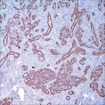
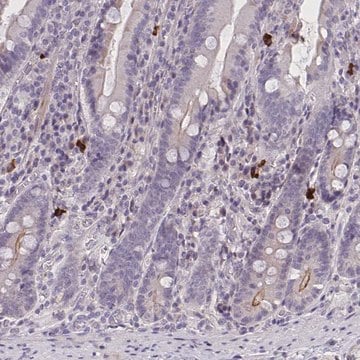
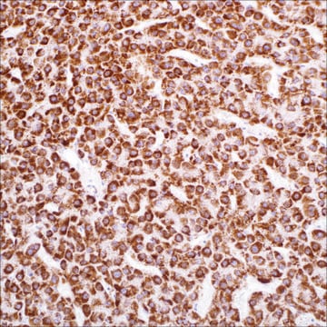
Gross cystic disease fluid protein-15 (GCDFP-15) is one of four major component proteins found in the cystic fluid obtained from patients with fibrocystic changes of the breast. GCDFP-15 is a marker of apocrine glandular differentiation in both benign and malignant mammary epithelium. This protein has a widespread distribution in apocrine glands elsewhere in the axillary and perianal tissues, as well as in the sublingual and submaxillary salivary glands. The GCDFP-15 protein is a 15 kDa glycoprotein shown to be prolactin-inducible, and the GCDFP-15 gene has been cloned. Ultrastructurally, the GCDFP-15 protein has been localized in Golgi vesicle and cytoplasmic granules. The protein is released by exocytosis at the apices of the mammary epithelial cells. The primary application for this antibody product is the identification of breast carcinoma in the metastatic setting, or fluid analysis. In a study by Wick et al. on paraffin sections of 105 breast cancers and 585 non-mammary malignancies in order to assess its value in this context, anti-GCDFP-15 was compared with another putative mammary epithelial marker, alpha-lactalbumin (ALA), with respect to sensitivity and specificity for a diagnosis of breast carcinoma. Overall, the rates of specificity and sensitivity and positive predictive value for GCDFP-15 were 95%, 74%, and 74%, respectively. A consistent congruency between the reactivity patterns of primary and metastatic breast cancers was noted for GCDFP-15. Besides mammary carcinomas, the major tumor types that express GCDFP-15 were carcinomas of the salivary glands and sweat glands. Since the latter two types of lesions are unlikely to be diagnosed as metastatic breast cancer, statistical indices were recalculated after exclusion of these three tumor types. Following this exclusion, the adjusted rate of specificity of GCDFP-15 and positive predictive value for a diagnosis of metastatic carcinoma of the breast were each 99%. Another study by Bhargava et al. has shown GCDFP-15 staining of a higher percentage of cancer cells in lobular carcinoma as compared to ductal carcinoma of the breast.

Liaison

GCDFP-15 Positive Control Slides, Product No. 257S, are available for immunohistochemistry (formalin-fixed, paraffin-embedded sections).

Forme physique

Solution in Tris Buffer, pH 7.3-7.7, with 1% BSA and <0.1% Sodium Azide

Notes préparatoires

Download the IFU specific to your product lot and formatNote: This requires a keycode which can be found on your packaging or product label.

Autres remarques

For Technical Service please contact: 800-665-7284 or email: service@cellmarque.com

Informations légales

Cell Marque is a trademark of Merck KGaA, Darmstadt, Germany

Vous ne trouvez pas le bon produit ?  

Essayez notre Outil de sélection de produits.

Code de la classe de stockage

12 - Non Combustible Liquids

Classe de danger pour l'eau (WGK)

WGK 2

Point d'éclair (°F)

Not applicable

Point d'éclair (°C)

Not applicable


Faites votre choix parmi les versions les plus récentes :

Certificats d'analyse (COA)

Lot/Batch Number

Vous ne trouvez pas la bonne version ?

Si vous avez besoin d'une version particulière, vous pouvez rechercher un certificat spécifique par le numéro de lot.

Déjà en possession de ce produit ?

Retrouvez la documentation relative aux produits que vous avez récemment achetés dans la Bibliothèque de documents.

Consulter la Bibliothèque de documents

G Mazoujian et al.
The American journal of pathology, 110(2), 105-112 (1983-02-01)
Gross cystic disease fluid is a pathologic secretion from breast composed of several glycoproteins, including a unique 15,000-dalton monomer protein, GCDFP-15. By the immunoperoxidase technique, GCDFP-15 was localized in the apocrine metaplastic epithelium lining breast cysts and in apocrine glands
S Ansai et al.
The American Journal of dermatopathology, 17(3), 249-255 (1995-06-01)
We investigated immunohistochemically the localization of lysozyme and Leu M1 in normal skin, 76 cases of benign sweat gland tumors, 28 cases of malignant sweat gland tumors, 23 cases of extramammary Paget's disease, 7 cases of sebaceous carcinoma, 6 cases
U Raju et al.
Modern pathology : an official journal of the United States and Canadian Academy of Pathology, Inc, 6(5), 516-520 (1993-09-01)
Signet ring carcinoma of the breast often metastasizes to gastrointestinal tract and female genital tract. We report clinicopathologic features of 10 breast carcinomas with signet ring features, five of which had unusual metastatic patterns. The primary breast tumor in all
G Mazoujian et al.
The American Journal of dermatopathology, 10(1), 28-35 (1988-02-01)
Sixty-five cases of benign sweat gland tumors of the skin were studied for the expression and localization of gross cystic disease fluid protein-15 (GCDFP-15) by immunoperoxidase methods. There was positive staining of tumors of probable apocrine differentiation in 10 of
C Cohen et al.
Archives of pathology & laboratory medicine, 117(3), 291-294 (1993-03-01)
Mammary Paget's disease has been said to result from epidermal spread by contiguity of primary intraductal carcinoma. To assess similar identity, we immunostained Paget's cells and underlying intraductal and/or invasive mammary carcinoma in 20 cases for cytokeratins, epithelial membrane antigen

Notre équipe de scientifiques dispose d'une expérience dans tous les secteurs de la recherche, notamment en sciences de la vie, science des matériaux, synthèse chimique, chromatographie, analyse et dans de nombreux autres domaines..

Contacter notre Service technique