Accéder au contenu
Merck
Toutes les photos(1)

Principaux documents

236M-9

Sigma-Aldrich

CEA (CEA31) Mouse Monoclonal Antibody

Se connecterpour consulter vos tarifs contractuels et ceux de votre entreprise/organisme


About This Item

Code UNSPSC :
12352203
Nomenclature NACRES :
NA.41

Source biologique

mouse

Niveau de qualité

100
500

Conjugué

unconjugated

Forme d'anticorps

culture supernatant

Type de produit anticorps

primary antibodies

Clone

CEA31, monoclonal

Description

For In Vitro Diagnostic Use in Select Regions (See Chart)

Forme

buffered aqueous solution

Espèces réactives

human

Conditionnement

vial of 0.1 mL concentrate (236M-94)
vial of 0.5 mL concentrate (236M-95)
bottle of 1.0 mL predilute (236M-97)
vial of 1.0 mL concentrate (236M-96)
bottle of 7.0 mL predilute (236M-98)

Fabricant/nom de marque

Cell Marque

Technique(s)

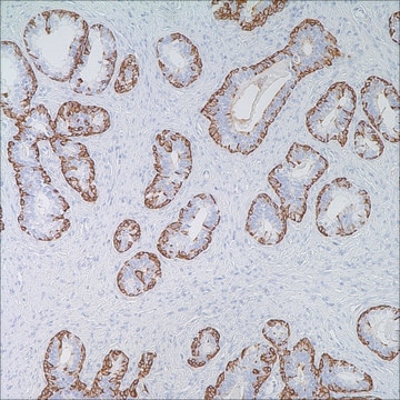
immunohistochemistry (formalin-fixed, paraffin-embedded sections): 1:100-1:500

Isotype

IgG1

Contrôle

colon

Conditions d'expédition

wet ice

Température de stockage

2-8°C

Visualisation

cytoplasmic

Informations sur le gène

human ... CEACAM5(1048)

Description générale

Anti-CEA is an antibody against carcinoembryonic antigen (CEA), a protein thought to promote tumor development through its role as a cell adhesion molecule. Anti-CEA positivity is seen in adenocarcinomas of many origins, especially colon and lung adenocarcinomas, but is rarely seen in mesothelial cells and mesotheliomas.

Qualité


IVD

IVD

IVD

RUO

Liaison

CEA Positive Control Slides, Product No. 236S, are available for immunohistochemistry (formalin-fixed, paraffin-embedded sections).

Forme physique

Solution in Tris Buffer, pH 7.3-7.7, with 1% BSA and <0.1% Sodium Azide

Notes préparatoires

Download the IFU specific to your product lot and formatNote: This requires a keycode which can be found on your packaging or product label.

Autres remarques

For Technical Service please contact: 800-665-7284 or email: service@cellmarque.com

Informations légales

Cell Marque is a trademark of Merck KGaA, Darmstadt, Germany

Vous ne trouvez pas le bon produit ?  

Essayez notre Outil de sélection de produits.


Certificats d'analyse (COA)

Recherchez un Certificats d'analyse (COA) en saisissant le numéro de lot du produit. Les numéros de lot figurent sur l'étiquette du produit après les mots "Lot" ou "Batch".

Déjà en possession de ce produit ?

Retrouvez la documentation relative aux produits que vous avez récemment achetés dans la Bibliothèque de documents.

Consulter la Bibliothèque de documents

V L Go
Cancer, 37(1 suppl), 562-566 (1976-01-01)
Carcinoembryionic antigen (CEA) a glycoprotein extracted from colonic cancer tissue (beta-globulin electrophoretic mobility, sedimentation coefficient 7 to 8S, and mol wt approximately 200,000) can be detected and measured by radioimmunoassay. Clinical evaluations of CEA determination have given the following results:
Julu Bhatnagar et al.
Anticancer research, 22(3), 1849-1857 (2002-08-10)
Although the prognostic value of Carcinoembryonic antigen (CEA) in colorectal cancer follow-up is well known, CEA expression in esophageal cancer is not widely recognized and studies correlating tissue CEA expression in stomach cancers with tumor differentiation have yielded contradictory results.
H Kamino et al.
Cancer, 61(6), 1142-1148 (1988-03-15)
Forty nasopharyngeal carcinomas (NPC) were studied by immunohistochemistry using an antibody to involucrin and the following three keratin antibodies: (1) an antibody to low molecular weight keratin reactive with nonsquamous epithelium, (2) a high molecular weight keratin antibody reactive with
J H Lagendijk et al.
Journal of clinical pathology, 52(4), 283-290 (1999-09-04)
To discriminate between adenocarcinomas that are primary to the ovary and metastatic to the ovary, especially of colonic and breast origin, by immunohistochemistry, using stepwise discriminant analysis or a decision tree. 312 routinely processed, formalin fixed tissue specimens were used.
V Tron et al.
Archives of pathology & laboratory medicine, 111(3), 291-293 (1987-03-01)
Immunohistologic markers have been of considerable value in differentiating malignant mesothelioma from adenocarcinoma. Recently, staining for milk-fat globule (MFG) protein has been suggested as a useful diagnostic test for this separation, but subsequent reports have been conflicting, with some authors

Notre équipe de scientifiques dispose d'une expérience dans tous les secteurs de la recherche, notamment en sciences de la vie, science des matériaux, synthèse chimique, chromatographie, analyse et dans de nombreux autres domaines..

Contacter notre Service technique