Accéder au contenu
Merck
Toutes les photos(2)

Principaux documents

MABT1507

Sigma-Aldrich

Anti-Desmoglein 2 Antibody, clone 6D8

clone 6D8, from mouse

Synonyme(s) :

Cadherin family member 5, HDGC

Se connecterpour consulter vos tarifs contractuels et ceux de votre entreprise/organisme


About This Item

Code UNSPSC :
12352203
eCl@ss :
32160702
Nomenclature NACRES :
NA.41

Source biologique

mouse

Forme d'anticorps

purified immunoglobulin

Type de produit anticorps

primary antibodies

Clone

6D8, monoclonal

Espèces réactives

human

Conditionnement

antibody small pack of 25 μL

Technique(s)

flow cytometry: suitable
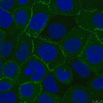
immunocytochemistry: suitable
immunoprecipitation (IP): suitable
inhibition assay: suitable
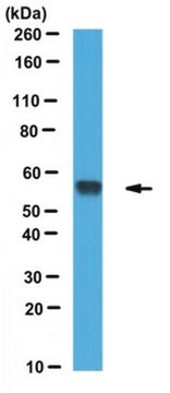
western blot: suitable

Isotype

IgG1κ

Numéro d'accès NCBI

Numéro d'accès UniProt

Modification post-traductionnelle de la cible

unmodified

Informations sur le gène

human ... DSG2(1829)

Description générale

Desmoglein-2 (UniProt: Q14126; also known as Cadherin family member 5, HDGC) is encoded by the DSG2 (also known as CDHF5) gene (Gene ID: 1829) in human. Desmogleins are protein substances located in and on the surface of keratinocytes. They are present in specialized cell-cell adhesion structures enriched in tissues that undergo substantial mechanical stress. These proteins have been shown to be a critical factor in cell-to-cell adhesion. Desmoglein-2 is a single-pass type I membrane protein that is found in all tissues and in carcinomas. It serves as a component of intercellular desmosome junctions. It is involved in the interaction of plaque proteins and intermediate filaments mediating cell-cell adhesion. It is synthesized with a signal peptide (aa 1-23) and a propeptide (aa 24-49) that are cleaved off in the mature form. It contains an extracellular domain (aa 50-609), a transmembrane domain (aa 610-634), and a cytoplasmic domain (aa 635-1118). Mutations in DSG2 gene have been linked to familial type of arrhythmogenic right ventricular dysplalsia that is characterized y infiltration of adipose and fibrous tissue into the right ventricle and loss of myocardial cells, resulting in ventricular and supraventricular arrhythmias.

Spécificité

Clone 6D8 specifically detects Desmoglein-2 in human cells.

Immunogène

Supernatant from cell membranes extract from A-431 cells.

Application

Anti-Desmoglein 2, clone 6D8, Cat. No. MABT1507, is a mouse monoclonal antibody that detects Desmoglein-2 and has been tested for use in Flow Cytometry, Immunocytochemistry, Immunoprecipitation, Inhibition assays, and Western Blotting,
Flow Cytometry Analysis: A representative lot detected Desmoglein 2 in Flow Cytometry applications (Tan, L.Y., et. al. (2016). Oncotarget. 7(29):46492-46508).

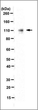
Western Blotting Analysis: A 1:125 dilution from a repesentative lot detected Desmoglein 2 in HaCaT cell lysates.

Flow Cytometry Analysis: 1 µg from a representative lot detected Desmoglein2 in SK-MEL-28.

Western Blotting Analysis: A representative lot detected Desmoglein 2 in Western Blotting applications (Wahl, J.K. 3rd, et. al. (2002). Hybrid Hybridomics. 21(1):37-44).

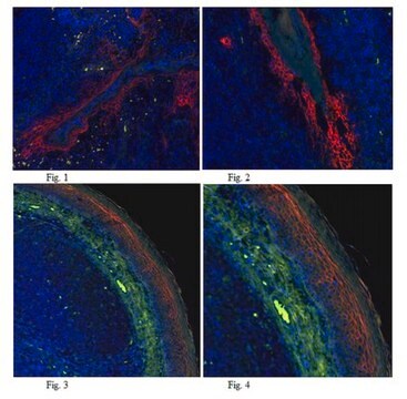
Immunocytochemistry Analysis: A representative lot detected Desmoglein 2 in Immunocytochemistry applications (Wahl, J.K. 3rd, et. al. (2002). Hybrid Hybridomics. 21(1):37-44; Tan, L.Y., et. al. (2016). Oncotarget. 7(29):46492-46508).

Immunoprecipitation Analysis: A representative lot immunoprecipitated Desmoglein 2 in Immunoprecipitation applications (Wahl, J.K. 3rd, et. al. (2002). Hybrid Hybridomics. 21(1):37-44).

Inhibition Analysis: A representative lotinhibited the attachment of adenovirus (Ad) serotypes Ad3.(Wang, H., et. al. (2011). Nat Med. 17(1):96-104).
Research Category
Cell Structure

Qualité

Evaluated by Immunocytochemistry in A431 cell line.

Immunocytochemistry Analysis: A 1:500 dilution of this antibody detected Desmoglein 2 in A431 cell line.

Description de la cible

~165 kDa observed; 122.29 kDa calculated. Uncharacterized bands may be observed in some lysate(s).

Forme physique

Format: Purified
Protein G purified
Purified mouse monoclonal antibody IgG1 in PBS without azide.

Stockage et stabilité

Stable for 1 year at -20°C from date of receipt. Handling Recommendations: Upon receipt and prior to removing the cap, centrifuge the vial and gently mix the solution. Aliquot into microcentrifuge tubes and store at -20°C. Avoid repeated freeze/thaw cycles, which may damage IgG and affect product performance.

Autres remarques

Concentration: Please refer to lot specific datasheet.

Clause de non-responsabilité

Unless otherwise stated in our catalog or other company documentation accompanying the product(s), our products are intended for research use only and are not to be used for any other purpose, which includes but is not limited to, unauthorized commercial uses, in vitro diagnostic uses, ex vivo or in vivo therapeutic uses or any type of consumption or application to humans or animals.

Vous ne trouvez pas le bon produit ?  

Essayez notre Outil de sélection de produits.


Certificats d'analyse (COA)

Recherchez un Certificats d'analyse (COA) en saisissant le numéro de lot du produit. Les numéros de lot figurent sur l'étiquette du produit après les mots "Lot" ou "Batch".

Déjà en possession de ce produit ?

Retrouvez la documentation relative aux produits que vous avez récemment achetés dans la Bibliothèque de documents.

Consulter la Bibliothèque de documents

Notre équipe de scientifiques dispose d'une expérience dans tous les secteurs de la recherche, notamment en sciences de la vie, science des matériaux, synthèse chimique, chromatographie, analyse et dans de nombreux autres domaines..

Contacter notre Service technique