Accéder au contenu
Merck
Toutes les photos(1)

Principaux documents

MABN866

Sigma-Aldrich

Anti-Myocilin (NT) Antibody, clone 7.1

clone 7.1, from mouse

Synonyme(s) :

Myocilin, Myocilin 55 kDa subunit, Trabecular meshwork-induced glucocorticoid response protein, Myocilin, N-terminal fragment, Myocilin 20 kDa N-terminal fragment

Se connecterpour consulter vos tarifs contractuels et ceux de votre entreprise/organisme


About This Item

Code UNSPSC :
12352203
eCl@ss :
32160702
Nomenclature NACRES :
NA.41

Source biologique

mouse

Niveau de qualité

Forme d'anticorps

purified immunoglobulin

Type de produit anticorps

primary antibodies

Clone

7.1, monoclonal

Espèces réactives

human, mouse

Technique(s)

immunohistochemistry: suitable
western blot: suitable

Isotype

IgG1κ

Numéro d'accès NCBI

Numéro d'accès UniProt

Conditions d'expédition

wet ice

Modification post-traductionnelle de la cible

unmodified

Informations sur le gène

human ... MYOC(4653)

Description générale

Myocilin (mutated trabecular meshwork-induced glucocorticoid response protein) is encoded by the MYOC gene (also known as GLC1A, GPOA, JOAG, JOAG1, TIGR) in human (Entrez Gene ID 4653). Myocilin is produced as a 504 amino acid glycoprotein containing an N-terminal hydrophobic signal peptide sequence, a coiled-coiled leucine zipper domain, and a C-terminal domain with homology to olfactomedins. Myocilin is commonly identified as a 53/57 kDa doublet by gel electrophoresis, and a 66 kDa form of the protein is also reported. Myocilin is known to undergo intracellular endoproteolytic cleavage between Glu214 and Leu215, resulting in a 20 kDa N-terminal and a 35 kDa C-terminal fragment. Myocilin is identified as a Lingo-1 receptor ligand and MYOC gene mutations are linked to 10% of juvenile open-angle glaucoma cases and 3-4% of those with primary open-angle glaucoma (PMID 24732711 24741044, 24837143). Cat. No. MABN866, clone 7.1 is a mouse monoclonal antibody that recognizes an epitope within the N-terminal fragment (aa33-214) and is demonstrated to be suitable for Western blotting, immuncytochemistry, and immunhistochemistry applications. This clone is reactive toward both human and murine species. (PMID 18674535 & 23979599).

Immunogène

Epitope: N-terminal fragment (aa 33-214)
Recombinant protein corresponding to the N-terminal fragment (aa 33-214) of human Myocilin.

Application

Anti-Myocilin Antibody, clone 7.1 is an antibody against Myocilin for use in Western Blotting, Immunohistochemistry.
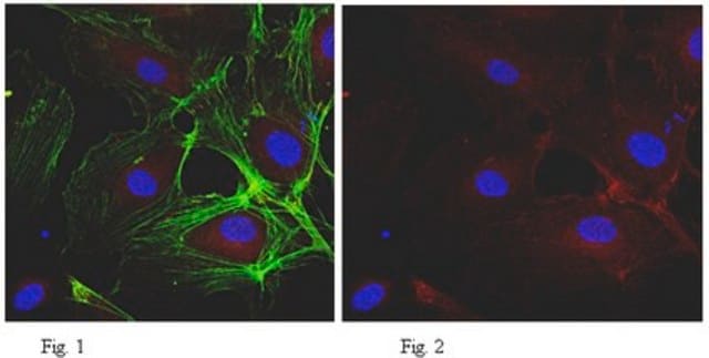
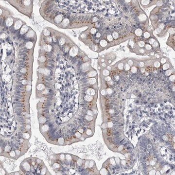
Immunohistochemistry Analysis: A representative lot detected Myocilin in human trabecular tissue (Ezzat, M.K., et al. (2008). Exp. Eye Res. 87(4):376-384).
Western Blotting Analysis: A representative lot detected Myocilin in human trabecular tissue (Ezzat, M.K., et al. (2008). Exp. Eye Res. 87(4):376-384).
Western Blotting Analysis: A representative lot detected Myocilin in aqueous humor (Zhao, Y., et al. (2013). Mol. Cell Biol. 33(21):4225-4240).
Research Category
Neuroscience
Research Sub Category
Developmental Signaling

Qualité

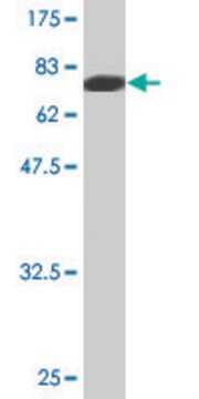
Evaluated by Western Blotting in mouse eye tissue lysate.

Western Blotting Analysis: 0.5 µg/mL of this antibody detected Myocilin in 10 µg of mouse eye tissue lysate.

Description de la cible

~53, 60 kDa observed

Forme physique

Format: Purified
Protein G Purified
Purified mouse monoclonal IgG1κ in buffer containing 0.1 M Tris-Glycine (pH 7.4), 150 mM NaCl with 0.05% sodium azide.

Stockage et stabilité

Stable for 1 year at 2-8°C from date of receipt.

Autres remarques

Concentration: Please refer to lot specific datasheet.

Clause de non-responsabilité

Unless otherwise stated in our catalog or other company documentation accompanying the product(s), our products are intended for research use only and are not to be used for any other purpose, which includes but is not limited to, unauthorized commercial uses, in vitro diagnostic uses, ex vivo or in vivo therapeutic uses or any type of consumption or application to humans or animals.

Vous ne trouvez pas le bon produit ?  

Essayez notre Outil de sélection de produits.

Code de la classe de stockage

12 - Non Combustible Liquids

Classe de danger pour l'eau (WGK)

WGK 1

Point d'éclair (°F)

Not applicable

Point d'éclair (°C)

Not applicable


Certificats d'analyse (COA)

Recherchez un Certificats d'analyse (COA) en saisissant le numéro de lot du produit. Les numéros de lot figurent sur l'étiquette du produit après les mots "Lot" ou "Batch".

Déjà en possession de ce produit ?

Retrouvez la documentation relative aux produits que vous avez récemment achetés dans la Bibliothèque de documents.

Consulter la Bibliothèque de documents

Biting Zhou et al.
Frontiers in genetics, 13, 1019208-1019208 (2022-10-22)
MYOC is a common pathogenic gene for primary open-angle glaucoma and encodes the protein named myocilin. Multiple MYOC variations have been found, with different clinical significance. However, the pathogenesis of glaucoma induced by MYOC mutations has not been fully clarified.
Maximilian Binter et al.
PloS one, 18(12), e0296124-e0296124 (2023-12-21)
The outflow pathway, especially trabecular meshwork (TM), plays an essential role in glaucoma, and the availability of TM cells is crucial for in vitro research. So far, the isolation of TM cells from mice has been anything but manageable due
Philip Mzyk et al.
International journal of molecular sciences, 23(4) (2022-02-27)
Although the extracellular matrix (ECM) in trabecular meshwork (TM) cells is known to be important in intraocular pressure (IOP) regulation, the molecular mechanisms involved in generating a glaucomatous environment in the TM are not completely understood. Recently we identified a
Sarah M Judge et al.
Cancer research, 80(9), 1861-1874 (2020-03-07)
Skeletal muscle wasting is a devastating consequence of cancer that contributes to increased complications and poor survival, but is not well understood at the molecular level. Herein, we investigated the role of Myocilin (Myoc), a skeletal muscle hypertrophy-promoting protein that
Kate E Keller et al.
Experimental eye research, 171, 164-173 (2018-03-13)
Cultured trabecular meshwork (TM) cells are a valuable model system to study the cellular mechanisms involved in the regulation of conventional outflow resistance and thus intraocular pressure; and their dysfunction resulting in ocular hypertension. In this review, we describe the

Notre équipe de scientifiques dispose d'une expérience dans tous les secteurs de la recherche, notamment en sciences de la vie, science des matériaux, synthèse chimique, chromatographie, analyse et dans de nombreux autres domaines..

Contacter notre Service technique