Accéder au contenu
Merck
Toutes les photos(1)

Principaux documents

MABF2119

Sigma-Aldrich

Anti-CD318 (CD6L) Antibody, clone 3A11

clone 3A11, from mouse

Synonyme(s) :

CUB domain-containing protein 1, Membrane glycoprotein gp140, Subtractive immunization M plus HEp3-associated 135 kDa protein, SIMA135, Transmembrane and associated with src kinases, CD6L

Se connecterpour consulter vos tarifs contractuels et ceux de votre entreprise/organisme


About This Item

Code UNSPSC :
12352203
eCl@ss :
32160702
Nomenclature NACRES :
NA.43

Source biologique

mouse

Forme d'anticorps

purified immunoglobulin

Type de produit anticorps

primary antibodies

Clone

3A11, monoclonal

Espèces réactives

human

Conditionnement

antibody small pack of 25 μg

Technique(s)

flow cytometry: suitable
immunocytochemistry: suitable
immunohistochemistry: suitable (paraffin)
immunoprecipitation (IP): suitable
western blot: suitable

Isotype

IgG1κ

Numéro d'accès NCBI

Numéro d'accès UniProt

Modification post-traductionnelle de la cible

unmodified

Informations sur le gène

human ... CDCP1(64866)

Description générale

CUB domain-containing protein 1 (UniProt: Q9H5V8; also known as Membrane glycoprotein gp140, Subtractive immunization M plus HEp3-associated 135 kDa protein, SIMA135, Transmembrane and associated with Src kinases, CD318, CD6L) is encoded by the CDCP1 (also known as TRASK) gene (Gene ID:64866) in human. CD318 is a single-pass membrane protein (isoform 1) that serves as a ligand for CD6. It is highly expressed in mitotic cells and has low expression during interphase. It is detected in high levels in skeletal muscle and colon tissue and in lower levels in kidney, placenta, lungs, and small intestine. Its levels are shown to be up-regulated in several human cancer tissues. It is also expressed in high levels in synovial tissues where it participates in CD6-dependent adhesion of T cells to synovial fibroblasts. Presence of CD318 on cancer cells is associated with their aggressiveness and metastatic potential. Preincubation of cancer cells with clone 3a11 is shown to enhance killing of these cells and inhibits their survival. CD318 is shown to be chemoattractive to T cells and levels of soluble CD318 are increased in the synovial fluid from patients with rheumatoid arthritis and juvenile inflammatory arthritis. CD318 is synthesized with a signal peptide (aa 1-29), which is cleaved off in the mature form. It has an extracellular domain (aa 30-667), a transmembrane domain (aa 668-688), and a cytoplasmic domain (aa 689-836). Three isoforms of CD318 have been described that are produced by alternative splicing. (Ref.: Ruth, JH., et al. (2021). JCI Insight. doi: 10.1172/jci.insight.145662. Enyindah-Asonye, G., et al. (2017). Proc. Natl. Acad. Sci. USA 114(33); E6912-E6921).

Spécificité

Clone 3A11 specifically detects CD318 in human cells. It targets an epitope with in the extracellular domain.

Immunogène

Epitope: extracellular domain
HBL-100 human mammary gland epithelial cells.

Application

Anti-CD318 (CD6L), clone 3A11, Cat. No. MABF2119, is a mouse monoclonal antibody that detects CD318 (CD6L) and has been tested for use in Flow Cytometry, Immunocytochemistry, Immunohistochemistry (Paraffin), Immunoprecipitation, and Western Blotting.
Immunoprecipitation Analysis: A representative lot immunoprecipitated CD318 (CD6L) in Immunoprecipitation applications (Saifullah, M.K., et. al. (2004). J Immunol. 173(10):6125-33; Enyindah-Asonye, G., et. al. (2017). Proc Natl Acad Sci U S A. 114(33):E6912-E6921).

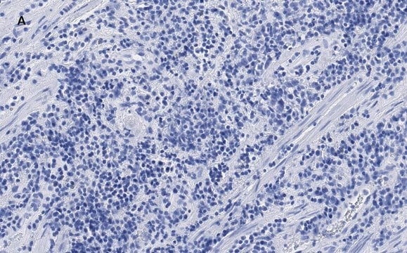
Immunohistochemistry (Paraffin) Analysis: A representative lot detected CD318 (CD6L) in Immunohistochemistry applications (Enyindah-Asonye, G., et. al. (2017). Proc Natl Acad Sci U S A. 114(33):E6912-E6921).

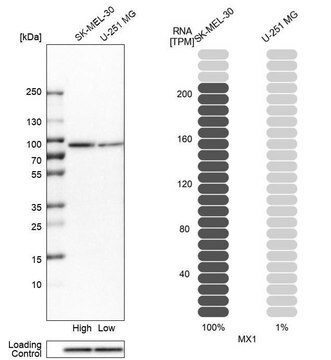
Western Blotting Analysis: A representative lot detected CD318 (CD6L) in Western Blotting applications (Saifullah, M.K., et. al. (2004). J Immunol. 173(10):6125-33; Enyindah-Asonye, G., et. al. (2017). Proc Natl Acad Sci U S A. 114(33):E6912-E6921).

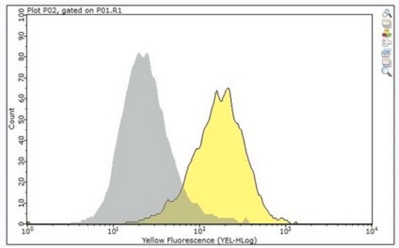
Flow Cytometry Analysis: A representative lot detected CD318 (CD6L) in Flow Cytometry applications (Saifullah, M.K., et. al. (2004). J Immunol. 173(10):6125-33; Enyindah-Asonye, G., et. al. (2017). Proc Natl Acad Sci U S A. 114(33):E6912-E6921).
Research Category
Inflammation & Immunology

Qualité

Evaluated by Immunocytochemistry in HaCaT epidermal keratinocyte cells.

Immunocytochemistry Analysis: A 1:1,000 dilution of this antibody detected CD318 (CD6L) in HaCaT epidermal keratinocyte cells.

Description de la cible

92.93 kDa calculated.

Forme physique

Format: Purified
Protein G purified
Purified mouse monoclonal antibody IgG1 in buffer containing 0.1 M Tris-Glycine (pH 7.4), 150 mM NaCl with 0.05% sodium azide.

Stockage et stabilité

Stable for 1 year at 2-8°C from date of receipt.

Autres remarques

Concentration: Please refer to lot specific datasheet.

Clause de non-responsabilité

Unless otherwise stated in our catalog or other company documentation accompanying the product(s), our products are intended for research use only and are not to be used for any other purpose, which includes but is not limited to, unauthorized commercial uses, in vitro diagnostic uses, ex vivo or in vivo therapeutic uses or any type of consumption or application to humans or animals.

Vous ne trouvez pas le bon produit ?  

Essayez notre Outil de sélection de produits.


Certificats d'analyse (COA)

Recherchez un Certificats d'analyse (COA) en saisissant le numéro de lot du produit. Les numéros de lot figurent sur l'étiquette du produit après les mots "Lot" ou "Batch".

Déjà en possession de ce produit ?

Retrouvez la documentation relative aux produits que vous avez récemment achetés dans la Bibliothèque de documents.

Consulter la Bibliothèque de documents

Notre équipe de scientifiques dispose d'une expérience dans tous les secteurs de la recherche, notamment en sciences de la vie, science des matériaux, synthèse chimique, chromatographie, analyse et dans de nombreux autres domaines..

Contacter notre Service technique