MAB3420
Anti-Tau-1 Antibody, clone PC1C6
clone PC1C6, Chemicon®, from mouse
Synonym(s):
Anti-Tau Antibody
Sign Into View Organizational & Contract Pricing
All Photos(2)
About This Item
UNSPSC Code:
12352203
eCl@ss:
32160702
NACRES:
NA.41
Recommended Products
biological source
mouse
Quality Level
antibody form
purified antibody
clone
PC1C6, monoclonal
species reactivity
human, rat, bovine
packaging
antibody small pack of 25 μg
manufacturer/tradename
Chemicon®
technique(s)
immunofluorescence: suitable
immunohistochemistry: suitable
western blot: suitable
isotype
IgG2a
NCBI accession no.
UniProt accession no.
shipped in
dry ice
storage temp.
−20°C
target post-translational modification
unmodified
General description
Tau, a microtubulebinding protein which serves to stabilize microtubules in growing axons, is found to be hyperphosphorylated in paired helical filaments (PHF), the major fibrous component of neurofibrillary lesions associated with Alzheimer’s disease. Hyperphosphorylation of Tau is thought to be the critical event leading to the assembly of PHF. Six Tau protein isoforms have been identified, all of which are phosphorylated by glycogen synthase kinase 3 (GSK 3). Cellular and subcellular localization: In situ, anti-tau-1 has a stringent specificity for the axons of neurons. The antibody does not stain the cell bodies or dendrites of neurons, nor does it stain any other cell type (4). However, this in vivo intracellular specificity is not maintained in culture: anti-tau-1 stains the axon, cell bodies, and dendrites of rat hippocampal neurons grown in culture (5). The specificity of anti-tau-1 was originally thought to represent the restricted expression of tau to axons. Later studies revealed that this specificity is dependant on the state of phosphorylation. In dephosphorylated samples (samples treated with alkaline phosphatase) anti-tau-1 stains astrocytes, perineuronal glial cells, and the axons, cell bodies and dendrites of neurons, while in untreated samples, anti-tau-1 stains only axons (6). (The epitope recognized by anti-tau-1 is probably at or near a phosphorylated site.)
Specificity
Anti-Tau-1 Antibody, clone PC1C6 binds to all known electrophoretic species of tau in human, rat and bovine brain (one-dimensional SDS-PAGE). However there is some unphosphorylated bias with clone PC1C6 as it seem to recognize only dephosphorylated serine sites at 195, 198, 199, and 202 {Szendrei, et al 1993; http://www.ncbi.nlm.nih.gov/entrez/query.fcgi-cmd=Retrieve&db=pubmed&dopt=Abstract&list_uids=7680727}. Also see Billingsley & Kincaid, 1997 Biochem J 323:577-591 for additional mapping information on PC1C6.
Immunogen
Purified denatured bovine microtubule associated proteins.
Application
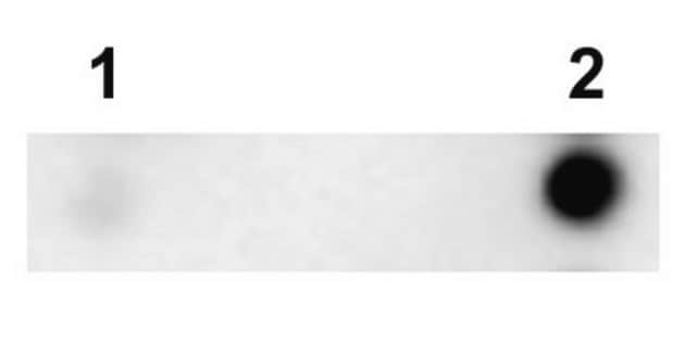
Anti-Tau-1 Antibody, clone PC1C6 is an antibody against Tau-1 for use in IH & WB with more than 65 product citations.
Research Category
Neuroscience
Neuroscience
Research Sub Category
Neurodegenerative Diseases
Neurodegenerative Diseases
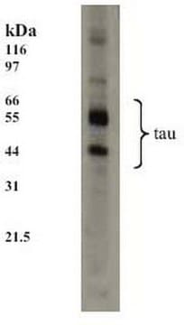
Western blot: Bovine brain microtubule proteins purified by two cycles of assembly and disassembly (9) are dissolved in SDS-PAGE sample buffer. Five micrograms of the microtuble preparation per lane is loaded onto a 4% to 20% SDS-PAGE gradient gel along side molecular weight markers (14.3 - 200 kD). After separation by electrophoresis, the proteins are blotted onto nitrocellulose. Tau is detected as a series of 5 bands (52-68 kD) with approximately 5 ng/mL of anti-tau1.
Immunofluorescence: A 1:1000 dilution of this antibody detected Tau in mouse primary neurons. (Basnet, N., et al. (2018). Nat. Cell Biol. 20(10); 1172-1180.
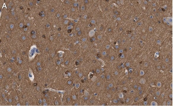
Immunohistochemistry: 5 μg/mL; stains axons in tissue primarily, however in culture Tau expression is not restricted to just axons.
Optimal working dilutions must be determined by end user.
Immunohistochemistry Protocol
Dephosphorylation of tissue sections (optional)
Dephosphorylation with alkaline phosphatase is recommended for staining neurofibrillary tangles in Alzheimer′s brain tissue with anti-tau-1 (6). This treatment changes the staining pattern of anti-tau-1 to include cell bodies, dendrites and axons of neurons. In untreated samples, anti-tau-1 stains axons only.
1. Incubate tissue sections at +32°C for 2.5 hours with constant agitation in the following solution: 100 mM Tris-HCl, pH 8.0; 130 units/mL alkaline phosphatase, 1 mM PMSF, 10 μg/mL pepstatin and 10 μg/mL leupeptin.
2. Rinse sections twice, 3 min per rinse, with 100 mM Tris-HCl, pH 8.0.
Anti-tau-1 staining
1. Block non-specific binding by incubating sections in PBS containing 1% (v/v) normal animal serum, and 0.03% (w/v) Triton X-100. The animal serum should be from the same species as the secondary antibody.
2. Rinse 3 times with PBS, 3 min per rinse.
3. Incubate sections with anti-tau-1, approximately 5 μg/mL, diluted in PBS containing 1% (v/v) normal animal serum.
4. Wash with PBS, changing the solution 3 times over a 3 min period.
5. Detect with a standard secondary antibody detection system (10-13).
Target description
5 bands (52–68 kDa)
Linkage
Replaces: AB1512
Physical form
0.02M phosphate buffer, pH 7.6, 0.25M NaCl, and 0.1% sodium azide
Format: Purified
Protein A purified
Storage and Stability
Maintain for 1 year at -20°C from date of shipment. Aliquot to avoid repeated freezing and thawing. For maximum recovery of product, centrifuge the original vial after thawing and prior to removing the cap.
Analysis Note
Control
Alzheimer′s brain tissue (dephosphorylation with alkaline phosphatase is recommended for staining neurofibrillary tangles in Alzheimer’s brain tissue) or human T98G glioblastoma cells
Alzheimer′s brain tissue (dephosphorylation with alkaline phosphatase is recommended for staining neurofibrillary tangles in Alzheimer’s brain tissue) or human T98G glioblastoma cells
Other Notes
Concentration: Please refer to the Certificate of Analysis for the lot-specific concentration.
Legal Information
CHEMICON is a registered trademark of Merck KGaA, Darmstadt, Germany
Disclaimer
Unless otherwise stated in our catalog or other company documentation accompanying the product(s), our products are intended for research use only and are not to be used for any other purpose, which includes but is not limited to, unauthorized commercial uses, in vitro diagnostic uses, ex vivo or in vivo therapeutic uses or any type of consumption or application to humans or animals.
recommended
Product No.
Description
Pricing
Storage Class Code
12 - Non Combustible Liquids
WGK
WGK 2
Flash Point(F)
Not applicable
Flash Point(C)
Not applicable
Certificates of Analysis (COA)
Search for Certificates of Analysis (COA) by entering the products Lot/Batch Number. Lot and Batch Numbers can be found on a product’s label following the words ‘Lot’ or ‘Batch’.
Already Own This Product?
Find documentation for the products that you have recently purchased in the Document Library.
Customers Also Viewed
Both the establishment and the maintenance of neuronal polarity require active mechanisms: critical roles of GSK-3beta and its upstream regulators.
Jiang, Hui, et al.
Cell, 120, 123-135 (2005)
Interaction of nonreceptor tyrosine-kinase Fer and p120 catenin is involved in neuronal polarization.
Lee, Seung-Hye
Molecules and Cells, 20, 256-262 (2005)
Katharina R L Schmitt et al.
Brain pathology (Zurich, Switzerland), 20(4), 771-779 (2010-01-15)
Systemic or brain-selective hypothermia is a well-established method for neuroprotection after brain trauma. There is increasing evidence that hypothermia exerts beneficial effects on the brain and may also support regenerative responses after brain damage. Here, we have investigated whether hypothermia
Centrosome motility is essential for initial axon formation in the neocortex.
de Anda, FC; Meletis, K; Ge, X; Rei, D; Tsai, LH
The Journal of Neuroscience null
Gradients of substrate-bound laminin orient axonal specification of neurons.
Dertinger, SK; Jiang, X; Li, Z; Murthy, VN; Whitesides, GM
Proceedings of the National Academy of Sciences of the USA null
Our team of scientists has experience in all areas of research including Life Science, Material Science, Chemical Synthesis, Chromatography, Analytical and many others.
Contact Technical Service


