Skip to Content
Merck
All Photos(2)

Key Documents

ABN363

Sigma-Aldrich

Anti-Myelin Protein P0 Antibody

from rabbit, purified by affinity chromatography

Synonym(s):

Myelin protein P0, Myelin peripheral protein, MPP, Myelin protein zero

Sign Into View Organizational & Contract Pricing


About This Item

UNSPSC Code:
12352203
eCl@ss:
32160702
NACRES:
NA.41

biological source

rabbit

Quality Level

antibody form

affinity isolated antibody

antibody product type

primary antibodies

clone

polyclonal

purified by

affinity chromatography

species reactivity

rat, mouse

species reactivity (predicted by homology)

bovine (based on 100% sequence homology), horse (based on 100% sequence homology), human (based on 100% sequence homology)

technique(s)

immunohistochemistry: suitable (paraffin)
western blot: suitable

NCBI accession no.

UniProt accession no.

shipped in

wet ice

target post-translational modification

unmodified

Gene Information

human ... MPZ(4359)

General description

Myelin protein P0 is also called Myelin peripheral protein (MPP) and Myelin protein zero (MPZ). Myelin protein P0 is found only in peripheral nervous system and is involved in the creation of an extracellular membrane face which guides the wrapping process that eventually leads to the compaction of adjacent lamellae. Myelin protein P0 is related to several disease states including Charcot-Marie-Tooth disease 1B (CMT1B), Charcot-Marie-Tooth disease 2I (CMT2I), Charcot-Marie-Tooth disease 2J (CMT2J), Adie pupil (ADIEP), Charcot-Marie-Tooth disease, dominant, intermediate type, D (CMTDID), Dejerine-Sottas syndrome (DSS), Congenital hypomyelination neuropathy (CHN), and Roussy-Levy syndrome (ROULS).

Immunogen

KLH-conjugated linear peptide corresponding to the extracellular domain of human Myelin protein P0.

Application

Anti-Myelin Protein P0 Antibody is a highly specific rabbit polyclonal antibody, that targets Myelin Protein & has been tested in western blotting & IHC (Paraffin).
Immunohistochemistry Analysis: A 1:2,000 dilution of this antibody detected Myelin protein P0 in rat spinal cord sections.
Research Category
Neuroscience
Research Sub Category
Developmental Neuroscience

Quality

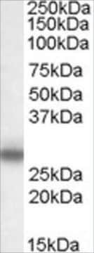
Evaluated by Western Blotting in rat spinal cord tissue lysate.

Western Blotting Analysis: A 1:5,000 dilution of this antibody detected Myelin protein P0 in 10 µg of rat spinal cord tissue lysate.

Target description

~30 kDa observed

Physical form

Affinity purified
Purified rabbit polyclonal in buffer containing 0.1 M Tris-Glycine (pH 7.4), 150 mM NaCl with 0.05% sodium azide.

Storage and Stability

Stable for 1 year at 2-8°C from date of receipt.

Disclaimer

Unless otherwise stated in our catalog or other company documentation accompanying the product(s), our products are intended for research use only and are not to be used for any other purpose, which includes but is not limited to, unauthorized commercial uses, in vitro diagnostic uses, ex vivo or in vivo therapeutic uses or any type of consumption or application to humans or animals.

Not finding the right product?  

Try our Product Selector Tool.

Storage Class Code

12 - Non Combustible Liquids

WGK

WGK 1

Flash Point(F)

Not applicable

Flash Point(C)

Not applicable


Certificates of Analysis (COA)

Search for Certificates of Analysis (COA) by entering the products Lot/Batch Number. Lot and Batch Numbers can be found on a product’s label following the words ‘Lot’ or ‘Batch’.

Already Own This Product?

Find documentation for the products that you have recently purchased in the Document Library.

Visit the Document Library

Ying Zou et al.
Cell & bioscience, 11(1), 210-210 (2021-12-16)
Silent information regulator 6 (SIRT6) is a mammalian homolog of the nicotinamide adenine dinucleotide (NAD)-dependent deacetylase sirtuin family. Prior evidences suggested that the anti-inflammatory function of SIRT6 after spinal cord and brain injury, and it plays a crucial role in
Ramazan Üstün et al.
Experimental and therapeutic medicine, 28(3), 345-345 (2024-07-29)
Traumatic and postoperative hemorrhages are life-threatening complications. Ankaferd BloodStopper (ABS) is a potent topical hemostatic agent to stop bleeding. However, ABS is associated with nerve toxicity. The present study aimed to investigate the functional and structural neurodegenerative effects of ABS
Sandra Murphy et al.
Journal of proteomics, 191, 212-227 (2018-02-07)
The highly progressive neuromuscular disorder dystrophinopathy is triggered by primary abnormalities in the Dmd gene, which causes cytoskeletal instability and loss of sarcolemmal integrity. Comparative organellar proteomics was employed to identify sarcolemma-associated proteins with an altered concentration in dystrophic muscle
Jiawei Xu et al.
Journal of neuroinflammation, 18(1), 234-234 (2021-10-17)
Plenty of macrophages are recruited to the injured nerve to play key roles in the immunoreaction and engulf the debris of degenerated axons and myelin during Wallerian degeneration, thus creating a conducive microenvironment for nerve regeneration. Recently, drugs targeting the
Yan Guo et al.
Signal transduction and targeted therapy, 9(1), 32-32 (2024-02-14)
The appropriate and specific response of nerve cells to various external cues is essential for the establishment and maintenance of neural circuits, and this process requires the proper recruitment of adaptor molecules to selectively activate downstream pathways. Here, we identified

Our team of scientists has experience in all areas of research including Life Science, Material Science, Chemical Synthesis, Chromatography, Analytical and many others.

Contact Technical Service