Skip to Content
Merck
All Photos(2)

Key Documents

ABF32

Sigma-Aldrich

Anti-Fibroblast-specific Protein 1 (S100A4) Antibody

from rabbit, purified by affinity chromatography

Synonym(s):

Protein S100-A4, Calvasculin, Metastasin, Placental calcium-binding protein, Protein Mts1, S100 calcium-binding protein A4

Sign Into View Organizational & Contract Pricing


About This Item

UNSPSC Code:
12352203
eCl@ss:
32160702
NACRES:
NA.41

biological source

rabbit

Quality Level

antibody form

affinity isolated antibody

antibody product type

primary antibodies

clone

polyclonal

purified by

affinity chromatography

species reactivity

mouse, rat, human, horse

species reactivity (predicted by homology)

canine (based on 100% sequence homology), bovine (based on 100% sequence homology), equine (based on 100% sequence homology)

technique(s)

immunocytochemistry: suitable
western blot: suitable

NCBI accession no.

UniProt accession no.

shipped in

wet ice

target post-translational modification

unmodified

Gene Information

human ... S100A4(6275)

General description

S100A4, also known as Fibroblast-specific Protein 1, is a member of the Ca2+- dependent S100 family of proteins containing 2 EF-hand calcium-binding motifs. S100 is a metastasis factor that may function in motility, invasion, and tubulin polymerization. Increased expression of S100A4 has been associated with various types of cancer.

Specificity

This antibody recognizes S100A4 at the C-terminus.

Immunogen

Epitope: C-terminus
KLH-conjugated linear peptide corresponding to the C-terminus of human S100A4.

Application

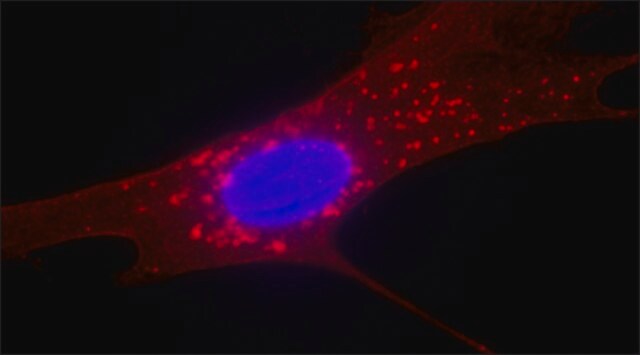
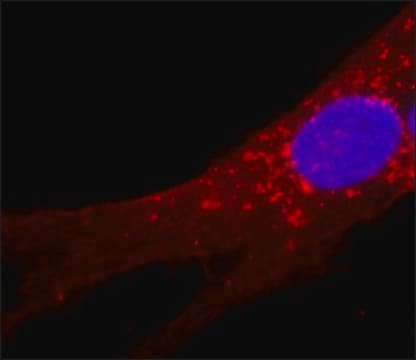
Immunocytochemistry Analysis: A 1:500 dilution from a representative lot detected S100A4 in mouse neural stem cells.
Research Category
Stem Cell Research

Inflammation & Immunology
Research Sub Category
Ion & Transport Channels

Inflammation & Autoimmune Mechanisms
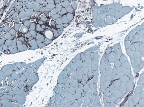
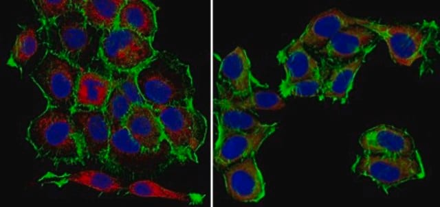
This Anti-Fibroblast-specific Protein 1 (S100A4) Antibody is validated for use in WB, IC for the detection of Fibroblast-specific Protein 1 (S100A4).

Quality

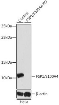
Evaluated by Western Blot in HeLa cell lysate.

Western Blot Analysis: 0.1 µg/mL of this antibody detected S100A4 in 10 µg of HeLa cell lysate.

Target description

~12 kDa observed

Physical form

Affinity purified
Purified rabbit polyclonal in buffer containing 0.1 M Tris-Glycine (pH 7.4), 150 mM NaCl with 0.05% sodium azide.

Storage and Stability

Stable for 1 year at 2-8°C from date of receipt.

Analysis Note

Control
HeLa cell lysate

Other Notes

Concentration: Please refer to the Certificate of Analysis for the lot-specific concentration.

Disclaimer

Unless otherwise stated in our catalog or other company documentation accompanying the product(s), our products are intended for research use only and are not to be used for any other purpose, which includes but is not limited to, unauthorized commercial uses, in vitro diagnostic uses, ex vivo or in vivo therapeutic uses or any type of consumption or application to humans or animals.

Not finding the right product?  

Try our Product Selector Tool.

Storage Class Code

12 - Non Combustible Liquids

WGK

WGK 1

Flash Point(F)

Not applicable

Flash Point(C)

Not applicable


Certificates of Analysis (COA)

Search for Certificates of Analysis (COA) by entering the products Lot/Batch Number. Lot and Batch Numbers can be found on a product’s label following the words ‘Lot’ or ‘Batch’.

Already Own This Product?

Find documentation for the products that you have recently purchased in the Document Library.

Visit the Document Library

Haiyang Tang et al.
American journal of physiology. Lung cellular and molecular physiology, 314(2), L256-L275 (2017-10-28)
Pulmonary vascular remodeling characterized by concentric wall thickening and intraluminal obliteration is a major contributor to the elevated pulmonary vascular resistance in patients with idiopathic pulmonary arterial hypertension (IPAH). Here we report that increased hypoxia-inducible factor 2α (HIF-2α) in lung
Nicole Y K Li-Jessen et al.
The Laryngoscope, 127(6), E193-E200 (2016-10-25)
High-mobility group box 1 (HMGB1) is a chromatin-binding protein located in the cell nucleus. Following injury, immunocompetent cells secrete HMGB1 to the extracellular milieu under the stimulation of proinflammatory cytokines. Extracellular HMGB1 acts a danger signal that instigates the innate
Carmelina Daniela Anfuso et al.
International journal of molecular sciences, 21(15) (2020-08-07)
Vascular pericytes are an important cellular component in the tumor microenvironment, however, their role in supporting cancer invasion is poorly understood. We hypothesized that PDGF-BB could be involved in the transition of human retinal pericytes (HRPC) in cancer-activated fibroblasts (CAF)
Juhi Pathak et al.
Scientific reports, 13(1), 13716-13716 (2023-08-23)
The enhanced availability of functional fibroblasts from precious tissue samples requires an ideal cell-culture system. Therefore, this study was designed to investigate the performance of caprine adult fibroblast cells (cadFibroblast) when cultivated in different culture media. The cadFibroblast cell lines
Liwei Jiang et al.
Cell, 183(5), 1219-1233 (2020-11-27)
Cancer therapies kill tumors either directly or indirectly by evoking immune responses and have been combined with varying levels of success. Here, we describe a paradigm to control cancer growth that is based on both direct tumor killing and the

Our team of scientists has experience in all areas of research including Life Science, Material Science, Chemical Synthesis, Chromatography, Analytical and many others.

Contact Technical Service