Direkt zum Inhalt
Merck

131M-9

Sigma-Aldrich

CD31 (JC70) Mouse Monoclonal Antibody

Anmeldenzur Ansicht organisationsspezifischer und vertraglich vereinbarter Preise


About This Item

UNSPSC-Code:
12352203
NACRES:
NA.41

Biologische Quelle

mouse

Qualitätsniveau

100
500

Konjugat

unconjugated

Antikörperform

culture supernatant

Antikörper-Produkttyp

primary antibodies

Klon

JC70, monoclonal

Beschreibung

For In Vitro Diagnostic Use in Select Regions (See Chart)

Form

buffered aqueous solution

Speziesreaktivität

human

Verpackung

vial of 0.1 mL concentrate (131M-94)
vial of 0.5 mL concentrate (131M-95)
bottle of 1.0 mL predilute (131M-97)
vial of 1.0 mL concentrate (131M-96)
bottle of 7.0 mL predilute (131M-98)

Hersteller/Markenname

Cell Marque

Methode(n)

immunohistochemistry (formalin-fixed, paraffin-embedded sections): 1:25-1:100

Isotyp

IgG1κ

Kontrolle

tonsil

Versandbedingung

wet ice

Lagertemp.

2-8°C

Visualisierung

cytoplasmic, membranous

Angaben zum Gen

human ... PECAM1(5175)

Allgemeine Beschreibung

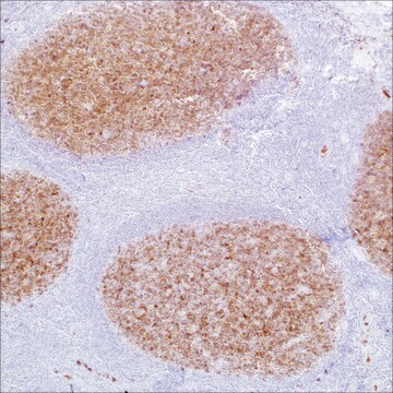
CD31, auch Thrombozyten-Endothelzellen-Adhäsionsmolekül (PECAM-1), ist ein vaskuläres Zelladhäsions-und Signalmolekül, das eine Rolle beim Erhalten der Gefäßpermeabilitätsbarrierean Endothelzellverbindungen spielt. Der immunhistochemische Nachweis von CD31 zeigt eine zytoplasmatischeund membranöse Expression von nichtneoplastischen und neoplastischen vaskulären Endothelzellen,einschließlich Neoplasien vaskulären Ursprungs wie Hämangiome und Angiosarkome.

Verlinkung

CD31-positive Kontrollobjektträger, Produktnr. 131S, sind für den Einsatz in der Immunhistochemie (formalinfixierte Paraffinschnitte) vorgesehen.

Physikalische Form

Lösung in Tris-Puffer, pH 7,3 - 7,7, mit 1 % BSA und <0,1 % Natriumazid

Angaben zur Herstellung

Laden Sie die für Ihre Produktcharge und Ihr Format geltende Gebrauchsanweisung herunterHinweis: Dies erfordert einen Schlüsselcode, der auf Ihrer Verpackung oder Ihrem Produktetikett angegeben ist.

Sonstige Hinweise

Den Technischen Kundendienst erreichen Sie unter: 800-665-7284 oder per E-Mail: service@cellmarque.com

Rechtliche Hinweise

Cell Marque is a trademark of Merck KGaA, Darmstadt, Germany

Sie haben nicht das passende Produkt gefunden?  

Probieren Sie unser Produkt-Auswahlhilfe. aus.

Lagerklassenschlüssel

12 - Non Combustible Liquids

WGK

WGK 2

Flammpunkt (°F)

Not applicable

Flammpunkt (°C)

Not applicable


Analysenzertifikate (COA)

Suchen Sie nach Analysenzertifikate (COA), indem Sie die Lot-/Chargennummer des Produkts eingeben. Lot- und Chargennummern sind auf dem Produktetikett hinter den Wörtern ‘Lot’ oder ‘Batch’ (Lot oder Charge) zu finden.

Besitzen Sie dieses Produkt bereits?

In der Dokumentenbibliothek finden Sie die Dokumentation zu den Produkten, die Sie kürzlich erworben haben.

Die Dokumentenbibliothek aufrufen

R L Attanoos et al.
Thorax, 55(10), 860-863 (2000-09-19)
Three cases of diffuse malignant vascular tumours of the pleura are described which mimicked malignant mesothelioma clinically and pathologically (so called "pseudomesothelioma"). All had occupational histories of exposure to asbestos. The relationship of these tumours to mesothelioma and asbestos exposure
Malak Abedalthagafi et al.
Annals of diagnostic pathology, 14(1), 15-22 (2010-02-04)
Cutaneous angiosarcoma (AS) is a rare malignant neoplasm of dermis composed of infiltrating cells of endothelial phenotype with overall poor prognosis. Although autocrine stimulation by vascular endothelial growth factor secretion may play a role in the pathogenesis of angiosarcoma, its
Xiu-Lian Xu et al.
The American Journal of dermatopathology, 32(8), 837-840 (2010-10-01)
Microvenular hemangioma (MVH) is an uncommon benign vascular neoplasm that usually occurs as a solitary asymptomatic red or purple papule, nodule, or plaque with a predilection for the upper extremities. Patients with more than 1 lesion, that is, multiple MVHs
CD31: An Immunospecific Marker for Endothelial Differentiation in Human Neoplasms
DeYoung BR, et al.
Appl. Immunohistochem., 1, 97-100 (1993)
F Alexander-Sefre et al.
Journal of clinical pathology, 56(10), 786-788 (2003-09-30)
Lymphovascular space invasion (LVSI) is an important step in the complex process of tumour metastasis. Various methods have been used in the past to improve the histological detection of LVSI. To develop a sensitive immunohistochemical method for the detection of

Unser Team von Wissenschaftlern verfügt über Erfahrung in allen Forschungsbereichen einschließlich Life Science, Materialwissenschaften, chemischer Synthese, Chromatographie, Analytik und vielen mehr..

Setzen Sie sich mit dem technischen Dienst in Verbindung.