Direkt zum Inhalt
Merck
Alle Fotos(3)

Wichtige Dokumente

MABN504

Sigma-Aldrich

Anti-VDAC1 Antibody, clone N152B/23

clone N152B/23, from mouse

Synonym(e):

Voltage-dependent anion-selective channel protein 1, VDAC-1, hVDAC1, Outer mitochondrial membrane protein porin 1, Plasmalemmal porin, Porin 31HL, Porin 31HM

Anmeldenzur Ansicht organisationsspezifischer und vertraglich vereinbarter Preise


About This Item

UNSPSC-Code:
12352203
eCl@ss:
32160702
NACRES:
NA.41

Biologische Quelle

mouse

Qualitätsniveau

Antikörperform

purified antibody

Antikörper-Produkttyp

primary antibodies

Klon

N152B/23, monoclonal

Speziesreaktivität

rat, mouse, human

Verpackung

antibody small pack of 25 μg

Methode(n)

immunohistochemistry: suitable
western blot: suitable

Isotyp

IgG2aκ

NCBI-Hinterlegungsnummer

UniProt-Hinterlegungsnummer

Versandbedingung

ambient

Lagertemp.

2-8°C

Posttranslationale Modifikation Target

unmodified

Angaben zum Gen

human ... VDAC1(7416)

Allgemeine Beschreibung

Voltage-dependent anion-selective channel protein 1 (VDAC1, VDAC), also known as outer mitochondrial membrane protein porin 1, is the outer mitochondrial membrane receptor for hexokinase and BCL2L1. VDAC1 forms a channel through the mitochondrial membrane and is involved in small molecule diffusion, cell volume regulation and apoptosis. VDAC1 may participate in the formation of the permeability transition pore complex (PTPC), which is responsible for the release of mitochondrial products that triggers apoptosis.

Spezifität

This antibody does not cross-react with VDAC2 or VDAC3 (Prof. J. Trimmer, University of California, Davis).

Immunogen

Recombinant protein corresponding to human VDAC1.

Anwendung

Research Category
Neurowissenschaft
Research Sub Category
Entwicklungsabhängige Signalübertragung
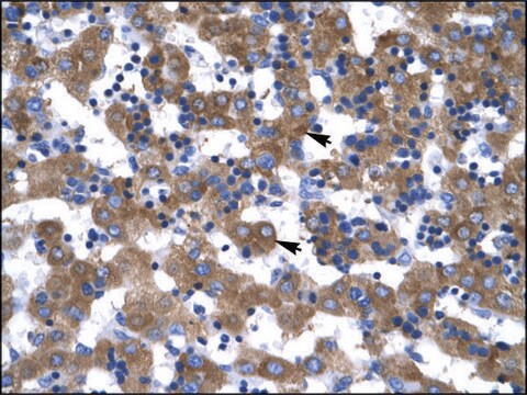
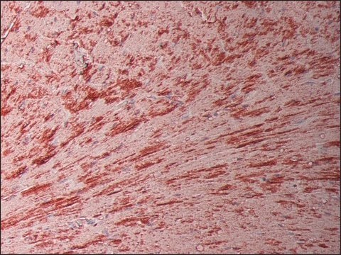
Detect VDAC using this mouse monoclonal antibody, Anti-VDAC1 Antibody, clone N152B/23 validated for use in western blotting & IHC.
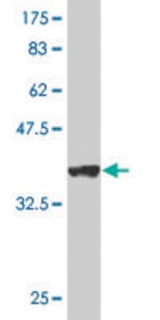
Western Blotting Analysis: A representative lot states VDAC1 does not cross-react with VDAC2 or VDAC3 (based on KO validation results) representative lot deteted VDAC1 in wild type mouse liver tissue, and demonstrated a loss of signal in VDAC1 knock out mouse hippocampal tissue using immunobloting analysis (Prof. J. Trimmer, University of California, Davis).
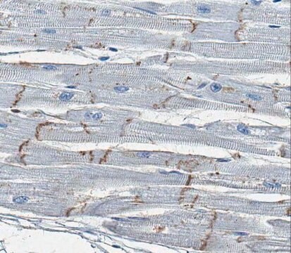
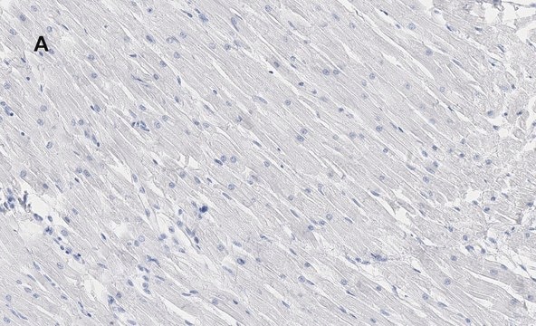
Immunohistochemistry Analysis: A 1:250 dilution from a representative lot detected VDAC1 in human cardiac myocytes tissue.

Qualität

Evaluated by Western Blotting in mouse brain tissue lysate.

Western Blotting Analysis: 0.5 µg/mL of this antibody detected VDAC1 in 10 µg of mouse brain tissue lysate.

Zielbeschreibung

~ 35 kDa observed

Physikalische Form

Protein G Purified
Format: Purified
Purified mouse monoclonal IgG2aκ in buffer containing 0.1 M Tris-Glycine (pH 7.4), 150 mM NaCl with 0.05% sodium azide.

Lagerung und Haltbarkeit

Stable for 1 year at 2-8°C from date of receipt.

Hinweis zur Analyse

Control
Mouse brain tissue lysate

Sonstige Hinweise

Concentration: Please refer to the Certificate of Analysis for the lot-specific concentration.

Haftungsausschluss

Unless otherwise stated in our catalog or other company documentation accompanying the product(s), our products are intended for research use only and are not to be used for any other purpose, which includes but is not limited to, unauthorized commercial uses, in vitro diagnostic uses, ex vivo or in vivo therapeutic uses or any type of consumption or application to humans or animals.

Sie haben nicht das passende Produkt gefunden?  

Probieren Sie unser Produkt-Auswahlhilfe. aus.

Empfehlung

Lagerklassenschlüssel

12 - Non Combustible Liquids

WGK

WGK 1

Flammpunkt (°F)

Not applicable

Flammpunkt (°C)

Not applicable


Analysenzertifikate (COA)

Suchen Sie nach Analysenzertifikate (COA), indem Sie die Lot-/Chargennummer des Produkts eingeben. Lot- und Chargennummern sind auf dem Produktetikett hinter den Wörtern ‘Lot’ oder ‘Batch’ (Lot oder Charge) zu finden.

Besitzen Sie dieses Produkt bereits?

In der Dokumentenbibliothek finden Sie die Dokumentation zu den Produkten, die Sie kürzlich erworben haben.

Die Dokumentenbibliothek aufrufen

Maria Feola et al.
Communications biology, 4(1), 517-517 (2021-05-05)
Erythropoiesis involves complex interrelated molecular signals influencing cell survival, differentiation, and enucleation. Diseases associated with ineffective erythropoiesis, such as β-thalassemias, exhibit erythroid expansion and defective enucleation. Clear mechanistic determinants of what make erythropoiesis effective are lacking. We previously demonstrated that
Mitofilin Heterozygote Mice Display an Increase in Myocardial Injury and Inflammation after Ischemia/Reperfusion.
Feng, et al.
Antioxidants (Basel, Switzerland), 12 (2023)
Ngonidzashe B Madungwe et al.
American journal of translational research, 12(7), 3412-3428 (2020-08-11)
MPV17 is an inner mitochondrial membrane protein whose mutation results in mitochondrial DNA (mtDNA) depletion diseases such as neurohepatopathy. MPV17 is expressed in several organs including the liver and kidneys. Here, we investigated its role and mechanism of action in
Chong Kiat Goo et al.
PloS one, 7(9), e45806-e45806 (2012-10-11)
Akt, a serine/threonine kinase has been shown to stimulate glycolysis in cancer cells but its role in mitochondrial respiration is unknown. Using PTEN-knockout mouse embryonic fibroblasts (MEF(PTEN-/-)) with hyper-activated Akt as a cell model, we observed a higher respiratory capacity
Abdulhafiz D Imam Aliagan et al.
American journal of translational research, 12(11), 7542-7564 (2020-12-15)
Mitochondrial dysfunction plays a critical role in the pathophysiology of Parkinson's disease (PD). The inner mitochondrial membrane (IMM) protein, Mitofilin or Mic60, has been shown to play a key role in controlling and maintaining mitochondrial cristae morphology, and its dysregulation

Unser Team von Wissenschaftlern verfügt über Erfahrung in allen Forschungsbereichen einschließlich Life Science, Materialwissenschaften, chemischer Synthese, Chromatographie, Analytik und vielen mehr..

Setzen Sie sich mit dem technischen Dienst in Verbindung.