Skip to Content
Merck
All Photos(2)

Documents

MABN119

Sigma-Aldrich

Anti-KLF6 Antibody, clone 12A8.3

clone 12A8.3, from mouse

Synonym(s):

Krueppel-like factor 6, B-cell-derived protein 1, Core promoter element-binding protein, GC-rich sites-binding factor GBF, Proto-oncogene BCD1, Suppressor of tumorigenicity 12 protein, Transcription factor Zf9

Sign Into View Organizational & Contract Pricing


About This Item

UNSPSC Code:
12352203
eCl@ss:
32160702
NACRES:
NA.41

biological source

mouse

Quality Level

antibody form

purified immunoglobulin

antibody product type

primary antibodies

clone

12A8.3, monoclonal

species reactivity

mouse, human

technique(s)

immunohistochemistry: suitable
western blot: suitable

isotype

IgG2bκ

NCBI accession no.

UniProt accession no.

shipped in

wet ice

target post-translational modification

unmodified

Gene Information

human ... KLF6(1316)

General description

KLF6 (Krueppel-like factor 6), is also known as BCD1, COPEB, CPBP and ST12. KLF6 contains zinc-finger domains and is thought to be a transcriptional activator binding to GC box motifs. KLF6 is involved in B-cell growth and development and has been shown to be highly expressed in placental cells as well as other tissues including the spleen, thymus, prostate, testis, small intestine and colon. KLF6 defects have been linked to gastric cancer (GASC) and prostate cancer (PC).

Immunogen

GST-tagged recombinant protein corresponding to human KLF6.

Application

Research Category
Neuroscience
Research Sub Category
Developmental Signaling
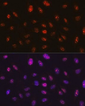
This KLF6 antibody is validated for use in WB & IHC for the detection of the KLF6 protein.
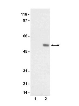
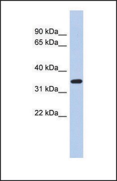
Western Blotting Analysis: 0.5 µg/mL from a representative lot detected KLF6 in 10 µg of human placenta tissue lysate.
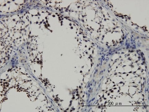
Immunohistochemistry Analysis: A 1:200 dilution from a representative lot detected KLF6 in human placenta tissue.

Quality

Evaluated by Western Blotting in NIH-3T3 cell lysate.

Western Blotting Analysis: 0.5 µg/mL of this antibody detected KLF6 in 10 µg of NIH-3T3 cell lysate.

Target description

~37 kDa observed. The calculated molecular weight is 32 kDa, however KLF6 has been shown at ~37 through ~40 kDa bands in western blots (Kimmeleman, A. C., et al. (2004). Oncogene. 23:5077-5083).

Physical form

Format: Purified
Protein G Purified
Purified mouse monoclonal IgG2bκ in buffer containing 0.1 M Tris-Glycine (pH 7.4), 150 mM NaCl with 0.05% sodium azide.

Storage and Stability

Stable for 1 year at 2-8°C from date of receipt.

Analysis Note

Control
NIH-3T3 cell lysate

Other Notes

Concentration: Please refer to the Certificate of Analysis for the lot-specific concentration.

Disclaimer

Unless otherwise stated in our catalog or other company documentation accompanying the product(s), our products are intended for research use only and are not to be used for any other purpose, which includes but is not limited to, unauthorized commercial uses, in vitro diagnostic uses, ex vivo or in vivo therapeutic uses or any type of consumption or application to humans or animals.

Not finding the right product?  

Try our Product Selector Tool.

Storage Class Code

12 - Non Combustible Liquids

WGK

WGK 1

Flash Point(F)

Not applicable

Flash Point(C)

Not applicable


Certificates of Analysis (COA)

Search for Certificates of Analysis (COA) by entering the products Lot/Batch Number. Lot and Batch Numbers can be found on a product’s label following the words ‘Lot’ or ‘Batch’.

Already Own This Product?

Find documentation for the products that you have recently purchased in the Document Library.

Visit the Document Library

Ireti Eni-Aganga et al.
The Journal of biological chemistry, 300(2), 105605-105605 (2024-01-02)
Prolidase (PEPD) is the only hydrolase that cleaves the dipeptides containing C-terminal proline or hydroxyproline-the rate-limiting step in collagen biosynthesis. However, the molecular regulation of prolidase expression remains largely unknown. In this study, we have identified overlapping binding sites for
Emmalee R Adelman et al.
Cancer discovery, 9(8), 1080-1101 (2019-05-16)
Aging is associated with functional decline of hematopoietic stem cells (HSC) as well as an increased risk of myeloid malignancies. We performed an integrative characterization of epigenomic and transcriptomic changes, including single-cell RNA sequencing, during normal human aging. Lineage-CD34+CD38- cells

Our team of scientists has experience in all areas of research including Life Science, Material Science, Chemical Synthesis, Chromatography, Analytical and many others.

Contact Technical Service