Skip to Content
Merck
All Photos(4)

Key Documents

AB1548

Sigma-Aldrich

Anti-NMDAR2A&B Antibody, pan

Chemicon®, from rabbit

Sign Into View Organizational & Contract Pricing


About This Item

UNSPSC Code:
12352203
eCl@ss:
32160702
NACRES:
NA.41

biological source

rabbit

Quality Level

antibody form

affinity isolated antibody

antibody product type

primary antibodies

clone

polyclonal

purified by

affinity chromatography

species reactivity

mouse, guinea pig, rat, human, rabbit, avian

manufacturer/tradename

Chemicon®

technique(s)

immunocytochemistry: suitable
immunohistochemistry (formalin-fixed, paraffin-embedded sections): suitable
immunoprecipitation (IP): suitable
western blot: suitable

NCBI accession no.

UniProt accession no.

shipped in

wet ice

target post-translational modification

unmodified

Gene Information

human ... GRIN2A(2903)

Specificity

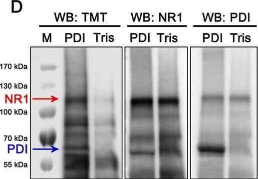
Western blot analysis of transfected cells shows the antibody recognizes both NR2A and NR2B subunits equally. There may be a very slight recognition of NR2C and NR2D. Western blot analysis of rat brain shows a single band comigrating with NMDAR2A and NMDAR2B expressed in transfected cells. AB1548 has also been used effectively in Western blot of the membrane fraction of rabbit cortex and avian brain homogenates. No cross-reaction is seen with NMDAR1 or other glutamate receptor subunits.

Immunogen

Synthetic peptide (LNSCSNRRVYKKMPSIESDV) corresponding to the C-terminus of rat NMDAR2A receptor subunit conjugated to BSA.

Application

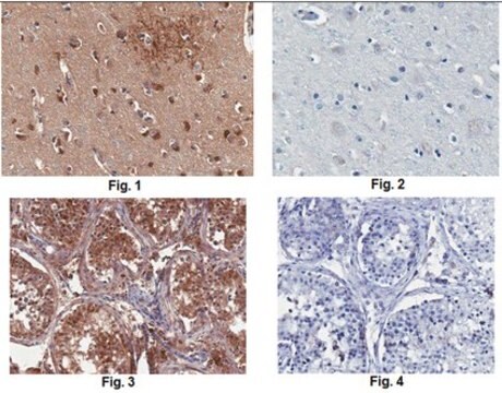
Anti-NMDAR2A&B Antibody, pan detects level of NMDAR2A&B & has been published & validated for use in IH(P), IC, IH, IP & WB.
Research Category
Neuroscience
Research Sub Category
Neurotransmitters & Receptors
Western blot analysis can be done at final concentrations of 0.25-0.5 μg/mL; recognizes a single band in membrane preparations of rat hippocampal neurons ~170,000-180,000 under reduced, SDS PAGE. AB1548 can be used for immunocytochemistry using paraformaldehyde or paraformaldehyde/glutaraldehyde fixed tissue with light and electron microscopy. Cryostat and vibratome sections can be used with or without Triton X-100 treatment. Final concentrations recommended are 0.5-1.0 μg/mL.

The antibody can be used for immunopreciptation of detergent solubilized receptor from brain or transfected cells. For most cases 5 μg of antibody is used with immobilized Protein A.

Optimal working dilutions must be determined by end user.

Physical form

Affinity purified using the immunogen peptide. Lyophilized from PBS containing 1% BSA.


Note: lot 0509010940 was shipped in liquid format; Do NOT add additional water to this lot of material.

Storage and Stability

Maintain lyophilized material at 2-8°C or -20°C. Reconstitute to 500 μL with sterile distilled water. Store reconstituted material frozen (-20°C or -80°C) in undiluted aliquots for up to six months. Avoid repeated thawing and refreezing. Diluted antibody containing 0.1% sodium azide for Western blot analysis can be stored for several weeks and reused repeatedly.

Legal Information

CHEMICON is a registered trademark of Merck KGaA, Darmstadt, Germany

Disclaimer

Unless otherwise stated in our catalog or other company documentation accompanying the product(s), our products are intended for research use only and are not to be used for any other purpose, which includes but is not limited to, unauthorized commercial uses, in vitro diagnostic uses, ex vivo or in vivo therapeutic uses or any type of consumption or application to humans or animals.

Not finding the right product?  

Try our Product Selector Tool.

Storage Class Code

10 - Combustible liquids


Certificates of Analysis (COA)

Search for Certificates of Analysis (COA) by entering the products Lot/Batch Number. Lot and Batch Numbers can be found on a product’s label following the words ‘Lot’ or ‘Batch’.

Already Own This Product?

Find documentation for the products that you have recently purchased in the Document Library.

Visit the Document Library

Grigory Krapivinsky et al.
Neuron, 40(4), 775-784 (2003-11-19)
The NMDA subtype of glutamate receptors (NMDAR) at excitatory neuronal synapses plays a key role in synaptic plasticity. The extracellular signal-regulated kinase (ERK1,2 or ERK) pathway is an essential component of NMDAR signal transduction controlling the neuroplasticity underlying memory processes
Vanessa Lage-Rupprecht et al.
eLife, 9 (2020-12-01)
In the rodent olfactory bulb the smooth dendrites of the principal glutamatergic mitral cells (MCs) form reciprocal dendrodendritic synapses with large spines on GABAergic granule cells (GC), where unitary release of glutamate can trigger postsynaptic local activation of voltage-gated Na+-channels
High prevalence of NMDA receptor IgA/IgM antibodies in different dementia types.
Doss, S; Wandinger, KP; Hyman, BT; Panzer, JA; Synofzik, M; Dickerson, B; Mollenhauer et al.
Annals of Clinical and Translational Neurology null
Integrins control dendritic spine plasticity in hippocampal neurons through NMDA receptor and Ca2+/calmodulin-dependent protein kinase II-mediated actin reorganization.
Shi, Y; Ethell, IM
The Journal of Neuroscience null
Multiple EphB receptor tyrosine kinases shape dendritic spines in the hippocampus.
Henkemeyer, M; Itkis, OS; Ngo, M; Hickmott, PW; Ethell, IM
The Journal of cell biology null

Our team of scientists has experience in all areas of research including Life Science, Material Science, Chemical Synthesis, Chromatography, Analytical and many others.

Contact Technical Service