Skip to Content
Merck
All Photos(3)

Documents

746223

Sigma-Aldrich

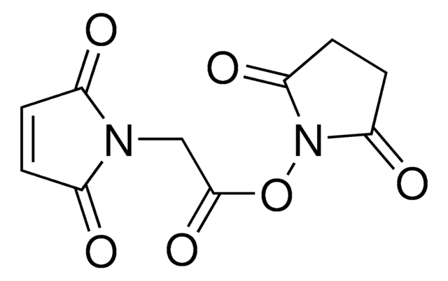
Maleimide-PEG2-succinimidyl ester

≥95%

Synonym(s):

3-[2-[2-[[3-(2,5-Dihydro-2,5-dioxo-1H-pyrrol-1-yl)-1-oxopropyl]amino]ethoxy]ethoxy]propanoic acid 2,5-dioxo-1-pyrrolidinyl ester, Maleimide-PEG2-NHS

Sign Into View Organizational & Contract Pricing


About This Item

Empirical Formula (Hill Notation):
C18H23N3O9
CAS Number:
Molecular Weight:
425.39
MDL number:
UNSPSC Code:
12352200
PubChem Substance ID:
NACRES:
NA.22

Assay

≥95%

form

solid

reaction suitability

reaction type: Pegylations
reagent type: cross-linking reagent

mp

92-94 °C

functional group

NHS ester
maleimide

storage temp.

−20°C

SMILES string

O=C(N1CCC(NCCOCCOCCC(ON(C(CC2)=O)C2=O)=O)=O)C=CC1=O

InChI

1S/C18H23N3O9/c22-13(5-8-20-14(23)1-2-15(20)24)19-7-10-29-12-11-28-9-6-18(27)30-21-16(25)3-4-17(21)26/h1-2H,3-12H2,(H,19,22)

InChI key

TZPDZOJURBVWHS-UHFFFAOYSA-N

Application

Heterobifunctional crosslinker with short ethylene oxide spacer for linking amine- to sulfhydryl-containing compounds or biomolecules. Maleimide functional group will react with sulfhydryls and the succinimidyl ester group will react with amines. Spacer length is 17.6 angstroms.

Storage Class Code

11 - Combustible Solids

WGK

WGK 3

Flash Point(F)

Not applicable

Flash Point(C)

Not applicable


Certificates of Analysis (COA)

Search for Certificates of Analysis (COA) by entering the products Lot/Batch Number. Lot and Batch Numbers can be found on a product’s label following the words ‘Lot’ or ‘Batch’.

Already Own This Product?

Find documentation for the products that you have recently purchased in the Document Library.

Visit the Document Library

Sinem K Saka et al.
Nature biotechnology, 37(9), 1080-1090 (2019-08-21)
Spatial mapping of proteins in tissues is hindered by limitations in multiplexing, sensitivity and throughput. Here we report immunostaining with signal amplification by exchange reaction (Immuno-SABER), which achieves highly multiplexed signal amplification via DNA-barcoded antibodies and orthogonal DNA concatemers generated
Sabrina Simoncelli et al.
Cell reports, 33(12), 108523-108523 (2020-12-29)
Elucidating the mechanisms that controlled T cell activation requires visualization of the spatial organization of multiple proteins on the submicron scale. Here, we use stoichiometrically accurate, multiplexed, single-molecule super-resolution microscopy (DNA-PAINT) to image the nanoscale spatial architecture of the primary inhibitor
Nirakar Basnet et al.
Nature cell biology, 20(10), 1172-1180 (2018-09-27)
Microtubules are central elements of the eukaryotic cytoskeleton that often function as part of branched networks. Current models for branching include nucleation of new microtubules from severed microtubule seeds or from γ-tubulin recruited to the side of a pre-existing microtubule.
Florian Schueder et al.
Nature communications, 8(1), 2090-2090 (2017-12-14)
Single-molecule localization microscopy (SMLM) can visualize biological targets on the nanoscale, but complex hardware is required to perform SMLM in thick samples. Here, we combine 3D DNA points accumulation for imaging in nanoscale topography (DNA-PAINT) with spinning disk confocal (SDC)
Thomas Schlichthaerle et al.
Chembiochem : a European journal of chemical biology, 20(8), 1032-1038 (2018-12-28)
Current optical super-resolution implementations are capable of resolving features spaced just a few nanometers apart. However, translating this spatial resolution to cellular targets is limited by the large size of traditionally employed primary and secondary antibody reagents. Recent advancements in

Our team of scientists has experience in all areas of research including Life Science, Material Science, Chemical Synthesis, Chromatography, Analytical and many others.

Contact Technical Service