Skip to Content
Merck
All Photos(2)

Key Documents

MABS132

Sigma-Aldrich

Anti-GLUT1 (CT) Antibody, clone 5B12.3

clone 5B12.3, from mouse

Synonym(s):

Solute carrier family 2 facilitated glucose transporter member 1, Glucose transporter type 1,erythrocyte/brain, GLUT-1, HepG2 glucose transporter

Sign Into View Organizational & Contract Pricing


About This Item

UNSPSC Code:
12352203
eCl@ss:
32160702
NACRES:
NA.41

biological source

mouse

Quality Level

antibody form

purified immunoglobulin

antibody product type

primary antibodies

clone

5B12.3, monoclonal

species reactivity

human, rat

packaging

antibody small pack of 25 μL

technique(s)

immunohistochemistry: suitable
western blot: suitable

isotype

IgG1κ

NCBI accession no.

UniProt accession no.

shipped in

ambient

storage temp.

2-8°C

target post-translational modification

unmodified

Gene Information

human ... SLC2A1(6513)

General description

GLUTs (Glucose transporters) are a family of integral membrane proteins that facilitative glucose uptake by cells. They are integral membrane glycoproteins involved in transporting glucose into most cells. Seven types of glucose transport carrier proteins, designated as GLUT 1 to 7, facilitate glucose transport across the cell membrane. Molecular cloning of glucose transporters have identified a family of closely related genes that encode at least 7 proteins exhibiting high degree of amino acid homology (45% to 65%), all in the molecular weight range of 40 to 60 kDa. GLUT1 functions in constitutive or basal glucose uptake in adult tissues and is the predominant glucose transporter in embryonic and fetal tissues. It has two forms, a 55 kDa and a 45 kDa. The 55 kDa form of GLUT1 regulates import of glucose from blood to brain across the endothelial cells of the blood-brain barrier (BBB), whereas the 45 kDa form of GLUT1 predominantly regulates nonvascular glial glucose uptake.

Specificity

This antibody recognizes GLUT1 at the C-terminus.

Immunogen

Epitope: C-terminus
KLH-conjugated linear peptide corresponding to the C-terminus of human GLUT1.

Application

Anti-GLUT1 (C-terminus) Antibody, clone 5B12.3 is an antibody against GLUT1 (C-terminus) for use in Western Blotting & IHC.
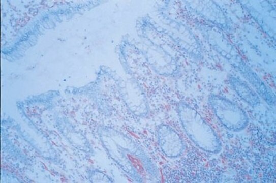
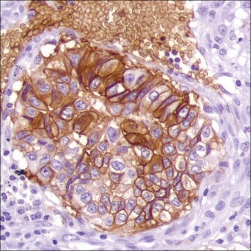
Immunohistochemistry Analysis: A 1:1,000 dilution from a representative lot detected GLUT1 in rat cerebellum and human cerebellum tissues.
Research Category
Signaling
Research Sub Category
Developmental Signaling

Quality

Evaluated by Western Blot in NGF treated PC-12 cell lysate.

Western Blot Analysis: 0.1 µg/mL of this antibody detected GLUT1 in 10 µg of NGF treated PC-12 cell lysate.

Target description

~54 kDa observed

Physical form

Format: Purified
Protein G Purified
Purified mouse monoclonal IgG1κ in buffer containing 0.1 M Tris-Glycine (pH 7.4), 150 mM NaCl with 0.05% sodium azide.

Storage and Stability

Stable for 1 year at 2-8°C from date of receipt.

Analysis Note

Control
NGF treated PC-12 cell lysate

Other Notes

Concentration: Please refer to the Certificate of Analysis for the lot-specific concentration.

Disclaimer

Unless otherwise stated in our catalog or other company documentation accompanying the product(s), our products are intended for research use only and are not to be used for any other purpose, which includes but is not limited to, unauthorized commercial uses, in vitro diagnostic uses, ex vivo or in vivo therapeutic uses or any type of consumption or application to humans or animals.

Not finding the right product?  

Try our Product Selector Tool.

Storage Class Code

12 - Non Combustible Liquids

WGK

WGK 1

Flash Point(F)

Not applicable

Flash Point(C)

Not applicable


Certificates of Analysis (COA)

Search for Certificates of Analysis (COA) by entering the products Lot/Batch Number. Lot and Batch Numbers can be found on a product’s label following the words ‘Lot’ or ‘Batch’.

Already Own This Product?

Find documentation for the products that you have recently purchased in the Document Library.

Visit the Document Library

Roberta Battistella et al.
International journal of molecular sciences, 22(21) (2021-11-14)
The vascular system is vital for all tissues and the interest in its visualization spans many fields. A number of different plant-derived lectins are used for detection of vasculature; however, studies performing direct comparison of the labeling efficacy of different
Stanley Andrisse et al.
American journal of physiology. Endocrinology and metabolism, 314(4), E353-E365 (2018-01-20)
Hyperandrogenemia and hyperinsulinemia are believed to play prominent roles in polycystic ovarian syndrome (PCOS). We explored the effects of low-dose dihydrotestosterone (DHT), a model of PCOS, on insulin signaling in metabolic and reproductive tissues in a female mouse model. Insulin
Linn Stenfelt et al.
Transfusion, 59(3), 1108-1117 (2019-01-01)
The P1 antigen was first described in 1927 and belongs to the P1PK histo-blood group system, together with Pk and NOR. The A4GALT-encoded 4-α-galactosyltransferase synthesizes these antigens and has been considered to extend glycolipids exclusively. However, contradicting studies have been
Jiaming Zhou et al.
ASN neuro, 15, 17590914231151534-17590914231151534 (2023-02-18)
Retinitis pigmentosa (RP) is an inherited disorder that results in vision impairment but general and mutation-independent therapeutic strategies are not available. However, it is widely regarded that the cGMP system, including cGMP and its interactor cGMP-dependent protein kinase (PKG), acts
PET Demonstrates Functional Recovery after Treatment by Danhong Injection in a Rat Model of Cerebral Ischemic-Reperfusion Injury.
Wang, Z; Song, F; Li, J; Zhang, Y; He, Y; Yang, J; Zhou, H; Zhao, T; Fu, W; Xing, P; Wan et al.
Evidence-Based Complementary and Alternative Medicine : ECAM null

Our team of scientists has experience in all areas of research including Life Science, Material Science, Chemical Synthesis, Chromatography, Analytical and many others.

Contact Technical Service