Skip to Content
Merck
All Photos(1)

Key Documents

810298C

Avanti

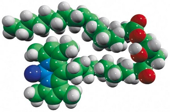
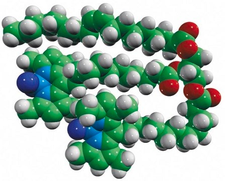
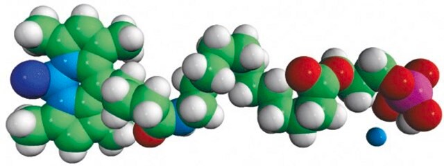
18:1-18:1-C11 TopFluor TG

Avanti Research - A Croda Brand 810298C

Synonym(s):

1,2-dioleoyl-3-[11-(dipyrrometheneboron difluoride)undecanoyl]-sn-glycerol

Sign Into View Organizational & Contract Pricing


About This Item

Empirical Formula (Hill Notation):
C63H105 BF2N2O6
CAS Number:
Molecular Weight:
1035.33
UNSPSC Code:
12352211
NACRES:
NA.25

Assay

>99% (TLC)

form

liquid

packaging

pkg of 1 × 1 mL (810298C-1mg)

manufacturer/tradename

Avanti Research - A Croda Brand 810298C

concentration

1 mg/mL (810298C-1mg)

shipped in

dry ice

storage temp.

−20°C

General description

Lipid droplets (LDs) are organelles that store lipids as reservoirs of metabolic energy and membrane lipid precursors, and they have been identified in the nucleus of some cells. LDs can be found in most eukaryotic cells. Most prominent are LDs in adipocytes, which make up most of the cellular volume, but other metabolically active cell types, such as liver or muscle cells, also have abundant LDs. LDs are unusual organelles in that they are bound by a monolayer of surface phospholipids, into which specific proteins are embedded, such as perilipins and metabolic enzymes. The cell biology of LDs as cellular organelles is only beginning to be unraveled. Avanti now offers a series of fluorescently-labeled mono-, di-, and triglycerides, which are useful for studying these important cellular organelles.

Application

18:1-18:1-C11TopFluor TG (1,2-dioleoyl-3-[11-(dipyrrometheneboron difluoride)undecanoyl]-sn-glycerol) has been used as a standard to validate fluorescent lipid bands in fatty acid incorporation assay. It may also be used in fluorescent lipid synthesis.

Packaging

5 mL Clear Glass Sealed Ampule (810298C-1mg)

Legal Information

Avanti Research is a trademark of Avanti Polar Lipids, LLC
TopFluor is a trademark of Avanti Polar Lipids, LLC

Pictograms

Skull and crossbonesHealth hazard

Signal Word

Danger

Hazard Classifications

Acute Tox. 3 Inhalation - Acute Tox. 4 Oral - Aquatic Chronic 3 - Carc. 2 - Eye Irrit. 2 - Repr. 2 - Skin Irrit. 2 - STOT RE 1 - STOT SE 3

Target Organs

Central nervous system, Liver,Kidney

WGK

WGK 3


Certificates of Analysis (COA)

Search for Certificates of Analysis (COA) by entering the products Lot/Batch Number. Lot and Batch Numbers can be found on a product’s label following the words ‘Lot’ or ‘Batch’.

Already Own This Product?

Find documentation for the products that you have recently purchased in the Document Library.

Visit the Document Library

Hanaa Hariri et al.
The Journal of cell biology, 218(4), 1319-1334 (2019-02-28)
Lipid droplets (LDs) serve as cytoplasmic reservoirs for energy-rich fatty acids (FAs) stored in the form of triacylglycerides (TAGs). During nutrient stress, yeast LDs cluster adjacent to the vacuole/lysosome, but how this LD accumulation is coordinated remains poorly understood. The
Øystein Sæle et al.
Biochimica et biophysica acta. Molecular and cell biology of lipids, 1863(9), 948-957 (2018-05-21)
The zebrafish larva is a powerful tool for the study of dietary triglyceride (TG) digestion and how fatty acids (FA) derived from dietary lipids are absorbed, metabolized and distributed to the body. While fluorescent FA analogues have enabled visualization of
Robert V Farese et al.
Cell, 139(5), 855-860 (2009-12-01)
Long underappreciated as important cellular organelles, lipid droplets are finally being recognized as dynamic structures with a complex and interesting biology. In light of this newfound respect, we discuss emerging views on lipid droplet biology and speculate on the major
Yuki Ohsaki et al.
The Journal of cell biology, 212(1), 29-38 (2016-01-06)
Lipid droplets (LDs) in the nucleus of hepatocyte-derived cell lines were found to be associated with premyelocytic leukemia (PML) nuclear bodies (NBs) and type I nucleoplasmic reticulum (NR) or the extension of the inner nuclear membrane. Knockdown of PML isoform
Robert V Farese et al.
The Journal of cell biology, 212(1), 7-8 (2016-01-06)
Lipid droplets (LDs) are sometimes found in the nucleus of some cells. In this issue, Ohsaki et al. (2016. J. Cell Biol. http://dx.doi.org/10.1083/jcb.201507122) show that the nuclear membrane, promyelocytic leukemia bodies, and the protein PML-II play a role in nuclear

Our team of scientists has experience in all areas of research including Life Science, Material Science, Chemical Synthesis, Chromatography, Analytical and many others.

Contact Technical Service