Lysosomal-associated membrane protein 1 (LAMP1), also termed LGP120, is a heavily glycosylated lysosomal membrane protein with a molecular mass of 120 kDa. It consists of a 40 kDa core polypeptide with O-linked and 18 asparagine-linked oligosaccharide side chains. LAMP1 protein contains a leader sequence, a large intralumenal region consisting of 2 homologous domains separated by a hinge region rich in proline and serine, a 24-amino acid transmembrane region, and a short cytoplasmic tail containing the lysosomal membrane targeting signal.
The gene LAMP1 (lysosomal-associated membrane protein 1) encodes a type I transmembrane protein that has a short cytoplasmic tail containing a lysosome-targeting signal of GYQTI(382)-COOH. The gene is mapped to human chromosome 13q34.
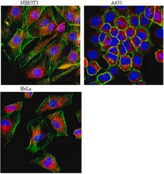
Application
Anti-LAMP1-Cy3™ antibody produced in rabbit has been used in immunofluorescence.[1][2]
Biochem/physiol Actions
The gene LAMP1 (lysosomal associated membrane protein 1) encodes a membrane glycoprotein that functions as an intracellular receptor. It is found to be expressed in the cytoplasm of several types of tumor cells and may be involved in tumor invasion. Lamp1 is crucial for perforin trafficking to the lytic granules and motility of these lytic granules. Its knockdown leads to inhibition of cytotoxicity of human natural killer cells.
Physical form
Solution in 0.01 M phosphate buffered saline, pH 7.4, containing 15 mM sodium azide.
Legal Information
Cy3 is a trademark of Cytiva
Disclaimer
Unless otherwise stated in our catalog or other company documentation accompanying the product(s), our products are intended for research use only and are not to be used for any other purpose, which includes but is not limited to, unauthorized commercial uses, in vitro diagnostic uses, ex vivo or in vivo therapeutic uses or any type of consumption or application to humans or animals.
Small (Weinheim an der Bergstrasse, Germany), 17(34), e2100887-e2100887 (2021-07-18)
The design of targeted nanomedicines requires intracellular space- and time-resolved data of nanoparticle distribution following uptake. Current methods to study intracellular trafficking, such as dynamic colocalization by fluorescence microscopy in live cells, are usually low throughput and require extensive analysis
It is demonstrated that repeated superovulation has deleterious effects on mouse ovaries and cumulus cells. However, little is known about the effects of repeated superovulation on early embryos. Epigenetic reprogramming is an important event in early embryonic development and could
European journal of immunology, 42(12), 3429-3441 (2012-09-29)
Podosomes, specialized actin-rich structures in macrophages (Mfs), degrade the extra-cellular matrix (ECM) and are involved in cell migration. On two-dimensional (2D) surfaces Mfs form spot-like podosomes at the ventral cell surface that develop into protrusive structures in a three-dimensional (3D)
Our team of scientists has experience in all areas of research including Life Science, Material Science, Chemical Synthesis, Chromatography, Analytical and many others.