dot blot: suitable immunocytochemistry: suitable indirect ELISA: suitable indirect immunofluorescence: 1:1,000 using human or other mammalian frozen sections western blot: suitable
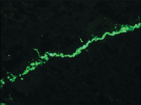
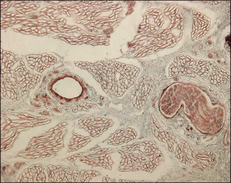
Collagen is a fibrous protein present in the extracellular framework of all vertebrates. Collagen type VII is the crucial component of anchoring fibrils and mutation in type VII collagen gene (COL7A1) leads to epidermolysis bullosa dystrophica. Monoclonal anti-collagen, type VII antibody can be used to distinguish invasive melanoma from non-invasive melanoma by visualizing the appearance and integrity of epidermal basement membrane. It can also be used in immunoblotting. Mouse anti-collagen type VII antibody reacts specifically with an epitope located on collagenase digested type VII collagen (150 kDa). The product also reacts with the basement membrane zone of stratified squamous epithelia of sheep, pig, bovine, guinea pig, goat and human.
Collagen type VII alpha 1 chain (COL7A1) is majorly expressed in skin, secreted by keratinocytes and fibroblasts. The gene is located on human chromosome 3p21.31.
Immunogen
insoluble fractions of neonatal human foreskin epidermal cells
Application
Monoclonal Anti-Collagen, Type VII antibody produced in mouse has been used in immuno-gold labelling.
Monoclonal anti-collagen type VII antibody can be used as a primary antibody in virus titration. It can also be used for the localization of type VII collagen in different immunochemical assays like dot blot, immunocytochemistry and ELISA.
Biochem/physiol Actions
Collagen type VII alpha 1 chain (COL7A1) is required for epithelium-to-stroma anchorage in skin, mucosa and cornea. It is considered as an immediate-early response gene for transforming growth factor β (TGF-β)/SMAD (mothers against decapentaplegic) signaling pathway.
Disclaimer
Unless otherwise stated in our catalog or other company documentation accompanying the product(s), our products are intended for research use only and are not to be used for any other purpose, which includes but is not limited to, unauthorized commercial uses, in vitro diagnostic uses, ex vivo or in vivo therapeutic uses or any type of consumption or application to humans or animals.
Molecular basis of dystrophic epidermolysis bullosa: mutations in the type VII collagen gene (COL7A1).
Jarvikallio A, Pulkkinen L
Human Mutation, 10(5), 338-347 (1997)
Type VII collagen associated with the basement membrane of amniotic epithelium forms giant anchoring rivets which penetrate a massive lamina reticularis
Efficient gene transfer into cultured fibroblasts and keratinocytes during retroviral transduction is a critical step toward the treatment of genodermatoses such as epidermolysis bullosa. However, achieving high transduction rates is still a difficult task, particularly for the insertion of large
SMAD3/4-dependent transcriptional activation of the human type VII collagen gene (COL7A1) promoter by transforming growth factor ?
Vindevoghel L, et al.
Proceedings of the National Academy of Sciences of the USA, 95(25), 14769-14774 (1998)
I would like to know the concentration of this antibody.
1 answer
Technical Support
·6 months ago
This product is a mouse ascites fluid. The concentration may range from 1.5 - 3.0 mg/mL and is reported in the lot specific Certificate of Analysis. Please see the link below to review a sample or lot specific Certificate: https://www.sigmaaldrich.com/product/sigma/c6805#product-documentation
Our team of scientists has experience in all areas of research including Life Science, Material Science, Chemical Synthesis, Chromatography, Analytical and many others.