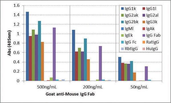
direct ELISA: 1:200,000 immunohistochemistry (formalin-fixed, paraffin-embedded sections): 1:300 western blot: 1:300,000-1:600,000 using using Hela cells extract
Immunoglobulin G (IgG) belongs to the immunoglobulin family and is a widely expressed serum antibody. Each immunoglobulin has two heavy chains and two light chains connected by a disulfide bond. It is a glycoprotein. IgG is a major class of immunoglobulin. Mouse consists of five immunoglobulin classes- IgM, IgG, IgA, IgD and IgE. Mouse IgG is further divided into five classes- IgG1, IgG2a, IgG2b and IgG3.
Application
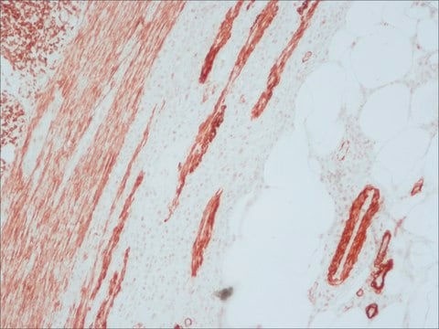
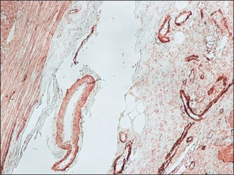
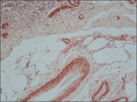
Anti-Mouse IgG (Fab specific)-Biotin antibody produced in goat has been used in immunohistochemistry and immunofluorescence studies.
Anti-Mouse IgG (Fab specific)-Biotin antibody produced in goat is suitable for direct ELISA at a dilution of 1:200000 and for immunohistochemistry at a dilution of 1:300. The antibody was used to stain paraffin-embedded, rat skin, duodenum, esophagus and spleen sections at a dilution of 1:200. For immunofluorescence of Human Gingival Fibroblasts, a dilution of 1:200 was used. For immunoblotting experiments using mouse adipose tissue, antibody dilution of 1:30000 was used.
Biochem/physiol Actions
IgG antibody subtype is the most abundant of serum immunoglobulins of the immune system. It is secreted by B cells and is found in blood and extracellular fluids and provides protection from infections caused by bacteria, fungi and viruses. Maternal IgG is transferred to fetus through the placenta that is vital for immune defense of the neonate against infections.
Immunoglobulin G (IgG) participates in hypersensitivity type II and type III reactions. IgG helps in opsonization, complement fixation and antibody dependent cell mediated cytotoxicity.
Other Notes
Antibody adsorbed with human IgG and rat serum proteins.
Physical form
Solution in 0.01 M phosphate buffered saline, pH 7.4, containing 15 mM sodium azide.
Preparation Note
Adsorbed to reduce background staining with human or rat samples.
Disclaimer
Unless otherwise stated in our catalog or other company documentation accompanying the product(s), our products are intended for research use only and are not to be used for any other purpose, which includes but is not limited to, unauthorized commercial uses, in vitro diagnostic uses, ex vivo or in vivo therapeutic uses or any type of consumption or application to humans or animals.
The journal of histochemistry and cytochemistry : official journal of the Histochemistry Society, 51(12), 1681-1688 (2003-11-19)
The standard method for assessment of cell proliferation in paraffin-embedded tissue sections is 5-bromodeoxyuridine (BrdU) immunohistochemistry (IHC). BrdU can be administered to laboratory animals via IP injections, is readily incorporated into nuclei during the DNA synthetic phase of the cell
Targeted disruption of fibronectin-integrin interactions in human gingival fibroblasts by the RI protease ofPorphyromonas gingivalis W50
Scragg MA, et al.
Infection and Immunity, 67(4), 1837-1843 (1999)
Dendrophthoe pentandra (L.) Miq extract effectively inhibits inflammation, proliferation and induces p53 expression on colitis-associated colon cancer
Endharti AT, et al.
BMC Complementary and Alternative Medicine, 16(1), 374-374 (2016)
There are indications that PRL may exert important metabolic actions on adipose tissue in different species. However, with the exception of birds, the receptor has not been identified in white adipose tissue. The present study was designed to examine the
Emerging high-field diffusion weighted MR imaging protocols, along with tractography, can elucidate microstructural changes associated with brain disease at the sub-millimeter image resolution. Epilepsy and other neurological disorders are accompanied by structural changes in the hippocampal formation and associated regions;
Our team of scientists has experience in all areas of research including Life Science, Material Science, Chemical Synthesis, Chromatography, Analytical and many others.