Skip to Content
Merck
All Photos(1)

Key Documents

M6190

Sigma-Aldrich

Monoclonal Anti-MYOD1 antibody produced in mouse

clone 5.2F, purified immunoglobulin, buffered aqueous solution

Synonym(s):

Anti-Myogenic Differentiation Antigen 1

Sign Into View Organizational & Contract Pricing


About This Item

MDL number:
UNSPSC Code:
12352203
NACRES:
NA.41

biological source

mouse

Quality Level

conjugate

unconjugated

antibody form

purified immunoglobulin

antibody product type

primary antibodies

clone

5.2F, monoclonal

form

buffered aqueous solution

mol wt

antigen 34 kDa

species reactivity

human, rat, chicken, mouse

concentration

1.0 mg/mL

technique(s)

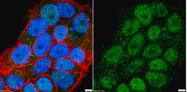
immunocytochemistry: suitable
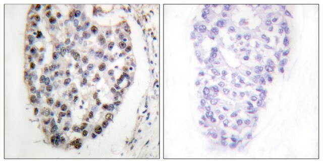
immunohistochemistry (formalin-fixed, paraffin-embedded sections): 2-4 μg/mL
immunohistochemistry (frozen sections): 2-4 μg/mL
immunoprecipitation (IP): 2 μg using 1 mg protein lysate
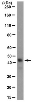
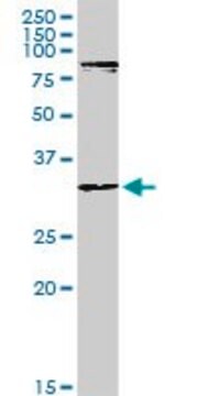
western blot: 1 μg/mL (reacts with the ~45 kDa protein)

isotype

IgG2a

UniProt accession no.

shipped in

wet ice

storage temp.

−20°C

Gene Information

human ... MYOD1(4654)
mouse ... Myod1(17927)
rat ... Myod1(337868)

General description

Myogenic differentiation antigen 1 (MYOD1) is a nuclear protein which is expressed in skeletal muscles. It is part of the basic helix-loop-helix (bHLH) family of transcription factors. The gene encoding it is localized on human chromosome 11.

Immunogen

recombinant mouse MyoD1 protein.

Application

Monoclonal Anti-MYOD1 antibody produced in mouse has been used in:
  • immunofluorescence staining at a 1:50 dilution
  • western blotting
  • immunostaining at a 1:300 dilution

Biochem/physiol Actions

Myogenic differentiation antigen 1 (MYOD1) maybe involved in recruitment of enzymes like acetyltransferases and methyltransferases to myogenic enhancers in the human genome. It takes part in the regeneration of muscles and mediates muscle cell differentiation by activating cell cycle arrest.

Physical form

Solution in phosphate buffered saline containing 0.08% sodium azide.

Disclaimer

Unless otherwise stated in our catalog or other company documentation accompanying the product(s), our products are intended for research use only and are not to be used for any other purpose, which includes but is not limited to, unauthorized commercial uses, in vitro diagnostic uses, ex vivo or in vivo therapeutic uses or any type of consumption or application to humans or animals.

Not finding the right product?  

Try our Product Selector Tool.

Storage Class Code

10 - Combustible liquids

WGK

nwg

Flash Point(F)

Not applicable

Flash Point(C)

Not applicable


Certificates of Analysis (COA)

Search for Certificates of Analysis (COA) by entering the products Lot/Batch Number. Lot and Batch Numbers can be found on a product’s label following the words ‘Lot’ or ‘Batch’.

Already Own This Product?

Find documentation for the products that you have recently purchased in the Document Library.

Visit the Document Library

Bolin Cai et al.
Cellular & molecular biology letters, 29(1), 9-9 (2024-01-05)
Skeletal muscle development is pivotal for animal growth and health. Recently, long noncoding RNAs (lncRNAs) were found to interact with chromatin through diverse roles. However, little is known about how lncRNAs act as chromatin-associated RNAs to regulate skeletal muscle development.
Shujie Chen et al.
Bio-protocol, 9(14), e3313-e3313 (2019-07-20)
Myofiber isolation followed with ex vivo culture could recapitulate and visualize satellite cells (SCs) activation, proliferation, and differentiation. This approach could be taken to understand the physiology of satellite cells and the molecular mechanism of regulatory factors, in terms of
Bahar Shahidi et al.
JOR spine, 3(2), e1087-e1087 (2020-07-03)
Many chronic musculoskeletal conditions are associated with loss of muscle volume and quality, resulting in functional decline. While atrophy has long been implicated as the mechanism of muscle loss in these conditions, recent evidence has emerged demonstrating a degenerative phenotype
Roy Blum
Journal of cellular biochemistry, 115(11), 1855-1867 (2014-06-07)
The early 1980s revelation of cis-acting genomic elements, known as transcriptional enhancers, is still regarded as one of the fundamental discoveries in the genomic field. However, only with the emergence of genome-wide techniques has the genuine biological scope of enhancers
Narasimhan P Agaram et al.
Genes, chromosomes & cancer, 53(9), 779-787 (2014-05-16)
Sclerosing and spindle cell rhabdomyosarcoma (RMS) are rare types of RMS recently reclassified as a stand-alone pathologic entity, separate from embryonal RMS (ERMS). Although sclerosing and spindle cell RMS share clinical and morphologic features, a pathogenetic link based on shared

Our team of scientists has experience in all areas of research including Life Science, Material Science, Chemical Synthesis, Chromatography, Analytical and many others.

Contact Technical Service