Skip to Content
Merck
All Photos(5)

Key Documents

G8918

Sigma-Aldrich

Anti-GRP78/BiP (GL-19) antibody produced in rabbit

enhanced validation

IgG fraction of antiserum, buffered aqueous solution

Synonym(s):

Anti-78-kDa Glucose-Regulated Protein, Anti-Immunoglobulin Heavy Chain Binding Protein

Sign Into View Organizational & Contract Pricing


About This Item

MDL number:
UNSPSC Code:
12352203
NACRES:
NA.41

biological source

rabbit

Quality Level

conjugate

unconjugated

antibody form

IgG fraction of antiserum

antibody product type

primary antibodies

clone

polyclonal

form

buffered aqueous solution

mol wt

antigen 78 kDa

species reactivity

mouse, hamster, human

enhanced validation

independent
Learn more about Antibody Enhanced Validation

technique(s)

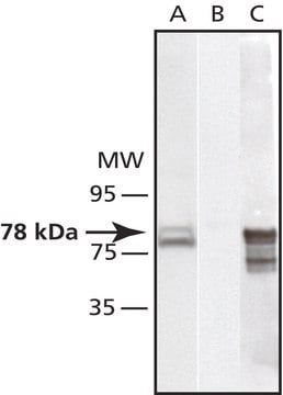
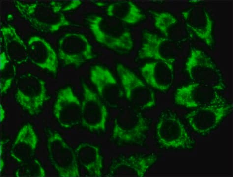
immunocytochemistry: 1:1,000 using mouse fibroblasts NIH3T3 cells
microarray: suitable
western blot: 1:3,000 using human epitheloid carcinoma HeLa whole cell extract

UniProt accession no.

shipped in

dry ice

storage temp.

−20°C

target post-translational modification

unmodified

Gene Information

human ... HSPA5(3309)
mouse ... Hspa5(14828)

General description

GRP78/BiP is a Ca2+-binding molecular chaperone located in the endoplasmic reticulum (ER). It is a highly conserved protein.

Application

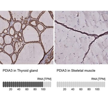
Anti-GRP78/BiP (GL-19) antibody produced in rabbit has been used in:
  • western blotting
  • immunocytochemistry
  • immunohistochemistry

Biochem/physiol Actions

GRP78 (78-kDa glucose-regulated protein) is essential for cell viability. GRP78/BiP is induced by stress-inducing agents or by conditions that adversely affect endoplasmic reticulum(ER) function, including oxidative stress, chemical toxicity, Ca2+ ionophores, inhibition of the ER Ca2+ -ATPase, inhibitors of glycosylation and hypoxia. GRP78/BiP is critical for maintenance of Ca2+ homeostasis and the prevention of apoptosis. GRP78/BiP levels are reduced in Alzheimer′s disease brains and the decreased expression of GRP78/BiP is found associated with mis-sense mutations in presenilin-1 (PS-1).

Physical form

Solution in 0.01 M phosphate buffered saline, pH 7.4, containing 15 mM sodium azide.

Disclaimer

Unless otherwise stated in our catalog or other company documentation accompanying the product(s), our products are intended for research use only and are not to be used for any other purpose, which includes but is not limited to, unauthorized commercial uses, in vitro diagnostic uses, ex vivo or in vivo therapeutic uses or any type of consumption or application to humans or animals.

Not finding the right product?  

Try our Product Selector Tool.

Storage Class Code

10 - Combustible liquids

WGK

nwg

Flash Point(F)

Not applicable

Flash Point(C)

Not applicable


Certificates of Analysis (COA)

Search for Certificates of Analysis (COA) by entering the products Lot/Batch Number. Lot and Batch Numbers can be found on a product’s label following the words ‘Lot’ or ‘Batch’.

Already Own This Product?

Find documentation for the products that you have recently purchased in the Document Library.

Visit the Document Library

miR-107 regulates granulin/progranulin with implications for traumatic brain injury and neurodegenerative disease
Wang WX, et al.
The American Journal of Pathology, 177(1), 334-345 (2010)
GRP78 at the centre of the stage in cancer and neuroprotection
Casas, Caty
Frontiers in Neuroscience, 11, 177-177 (2017)
Overexpressing GRP78 influences Ca2+ handling and function of mitochondria in astrocytes after ischemia-like stress
Ouyang YB, et al.
Mitochondrion, 11(2), 279-286 (2011)
Eva Ramon et al.
The Journal of biological chemistry, 289(52), 35918-35928 (2014-11-02)
Retinitis pigmentosa (RP) is a group of genetically and clinically heterogeneous inherited degenerative retinopathies caused by abnormalities of photoreceptors or retinal pigment epithelium in the retina leading to progressive sight loss. Rhodopsin is the prototypical G-protein-coupled receptor located in the
Henning G Hansen et al.
Bioscience reports, 34(2) (2014-04-01)
In the ER (endoplasmic reticulum) of human cells, disulfide bonds are predominantly generated by the two isoforms of Ero1 (ER oxidoreductin-1): Ero1α and Ero1β. The activity of Ero1α is tightly regulated through the formation of intramolecular disulfide bonds to help

Our team of scientists has experience in all areas of research including Life Science, Material Science, Chemical Synthesis, Chromatography, Analytical and many others.

Contact Technical Service