Skip to Content
Merck
All Photos(1)

Key Documents

G2279

Sigma-Aldrich

Monoclonal Anti-β-COP antibody produced in mouse

clone M3A5, ascites fluid

Synonym(s):

Anti-BARMACS, Anti-COPB

Sign Into View Organizational & Contract Pricing


About This Item

MDL number:
UNSPSC Code:
12352203
NACRES:
NA.41

biological source

mouse

Quality Level

conjugate

unconjugated

antibody form

ascites fluid

antibody product type

primary antibodies

clone

M3A5, monoclonal

contains

15 mM sodium azide

species reactivity

monkey, human, chicken, goose, rabbit, canine, bovine, kangaroo rat, rat, hamster

technique(s)

immunocytochemistry: suitable
immunoprecipitation (IP): suitable
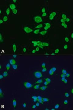
indirect immunofluorescence: 1:20 using cultured Chinese hamster ovary (CHO) cells

isotype

IgG1

UniProt accession no.

shipped in

dry ice

storage temp.

−20°C

target post-translational modification

unmodified

Gene Information

human ... COPB1(1315)
rat ... Copb1(114023)

General description

Monoclonal Anti- β-COP (mouse IgG1 isotype) is derived from the M3A5 hybridoma produced by the fusion of mouse myeloma cells and splenocytes from an immunized mouse. COPs (coatomer proteins) have a molar mass of 110 kDa and its primary structure is homologous to the β-adaptin component of clathrin-coated vesicles.
The coatomer (approx. 550kDa) consists of proteins designated α-, β-, γ-, and δ-COP, together with substoichiometric amounts of several other proteins.

Specificity

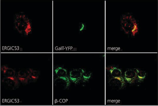
The antibody recognizes an epitope shared by β-COP (110 kDa) found in most tissue culture lines, and by a doublet of brain microtubule-associated protein (MAP2, 270-300 kDa). The antibody stains a reticular structure in the perinuclear area of non-neuronal cells (the periphery of Golgi complex and a population of coatomers scattered throughout the cytoplasm) in tissues from different species and cell processes, as well as cell bodies in chicken brain neuronal cells. It has been used for studies on the effects of various agents that influence energy status, disrupt the Golgi complex, or alter the activity of G-proteins or small GTP-binding proteins on the cellular localization of β-COP. The antibody may be used for the immunoaffinity purification of the protein.

Immunogen

microtubule-associated protein from goose brain.

Application

Monoclonal Anti-β-COP antibody produced in mouse has been used:
  • for the localization of β-COP using immunoprecipitation
  • in immunocytochemistry
  • in immunoblotting
  • with other antibodies to Golgi proteins to study the role and relationships of this protein in the cell

Biochem/physiol Actions

COPs (coatomer proteins) are transiently attached to the vesicles involved in transport within the Golgi complex and possibly between the rough ER and Golgi complex.

Disclaimer

Unless otherwise stated in our catalog or other company documentation accompanying the product(s), our products are intended for research use only and are not to be used for any other purpose, which includes but is not limited to, unauthorized commercial uses, in vitro diagnostic uses, ex vivo or in vivo therapeutic uses or any type of consumption or application to humans or animals.

Not finding the right product?  

Try our Product Selector Tool.

Storage Class Code

10 - Combustible liquids

WGK

nwg

Flash Point(F)

Not applicable

Flash Point(C)

Not applicable


Certificates of Analysis (COA)

Search for Certificates of Analysis (COA) by entering the products Lot/Batch Number. Lot and Batch Numbers can be found on a product’s label following the words ‘Lot’ or ‘Batch’.

Already Own This Product?

Find documentation for the products that you have recently purchased in the Document Library.

Visit the Document Library

J G Donaldson et al.
Science (New York, N.Y.), 254(5035), 1197-1199 (1991-11-22)
The binding of cytosolic coat proteins to organelles may regulate membrane structure and traffic. Evidence is presented that a small guanosine triphosphate (GTP)-binding protein, the adenosine diphosphate ribosylation factor (ARF), reversibly associates with the Golgi apparatus in an energy, GTP
J S Li et al.
Journal of virology, 70(9), 6029-6035 (1996-09-01)
Infection by human and animal hepadnaviruses displays remarkable host and tissue tropism. The infection cycle probably initiates with binding of the pre-S domain of viral envelope protein to surface receptors present on the hepatocyte. Three types of neutralizing monoclonal antibodies
G J Choukroun et al.
The Journal of clinical investigation, 106(8), 983-993 (2000-10-18)
The Golgi complex and the trans-Golgi network are critical cellular organelles involved in the endocytic and biosynthetic pathways of protein trafficking. Lipids have been implicated in the regulation of membrane-protein trafficking, vesicular fusion, and targeting. We have explored the role
T E Kreis et al.
Annual review of cell and developmental biology, 11, 677-706 (1995-01-01)
Cytosolic coat proteins (COPs) regulate membrane traffic in eukaryotic cells. Three classes of coat protein complexes have so far been identified: clathrin and its adaptor proteins, coatomer (COPI), and COPII. Coatomer (composed of seven different subunits) and ADP-ribosylation factor (ARF)
Cristina Torres et al.
Molecular microbiology, 54(3), 632-646 (2004-10-20)
Protozoan parasites are responsible of important healthy problems, among others malaria, leishmaniasis and trypanosomiasis. The present work reports the characterization of the first mammalian ATP-binding cassette transporter, subfamily A (ABCA)-like in Trypanosoma cruzi. TcABC1 is a single copy gene differentially

Our team of scientists has experience in all areas of research including Life Science, Material Science, Chemical Synthesis, Chromatography, Analytical and many others.

Contact Technical Service