General description
Severe Acute Respiratory Syndrome Coronavirus 2 (SARS-CoV-2) or (2019-nCoV) is a novel coronavirus that had spread on December 2019 in Hubei province of China and infected millions of people worldwide.1 The causative agent of COVID-19, the SARS-CoV-2 virus is a positive-strand RNA virus .The mature SARS-CoV-2 contains 4 structural proteins: Envelope (E), Membrane (M), Nucleocapsid (N), and the Spike protein (S), E and M proteins help in viral assembly and N protein is needed for RNA synthesis. The main receptor for SARS-CoV and SARS-CoV-2 on the membrane of the target cells is the Angiotensin 2 Converting Enzyme (ACE2), a metallopeptidase present on the membrane of many cells, including type-I and -II pneumocytes, small intestine enterocytes, kidney proximal tubules cells, the endothelial cells of arteries and veins, and the arterial smooth muscle, among other tissues.15-16 It has been shown that SARS-CoV-2 virus employs transmembrane protease serine 2 (TMPRSS2) for S protein priming and it is speculated that furin-mediated cleavage at the S1/S2 site in infected cells may promote subsequent TMPRSS2-dependent entry into target cells.
Specificity
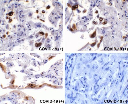
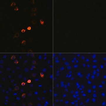
Anti-SARS-CoV-2-Spike protein C-terminal antibody specifically recognizes Spike from COVID-19 virus origin.
Immunogen
Synthetic peptide CFGD-15
Application
The antibody may be used in various immunochemical techniques including Immunoblotting and ELISA. Detection of the Spike RBD protein band by Immunoblotting is specifically inhibited by the immunogen.
Biochem/physiol Actions
The Spike protein (S) is responsible for virus binding and entry into host cells. The S precursor protein of SARS-CoV-2 is cleaved into S1 subunit (685 amino acids), and S2 (588 amino acids) subunits. S1 subunit has the receptor binding domain (RBD) that mediates entry of this virus into susceptible cells through the peptidase domain of host ACE2 with high affinity (Kd = 15 nM). S2 protein, which is reported to be well-conserved and shows 99% identity with bat coronavirus, is responsible for membrane fusion. The Spike protein is the most studied of the coronaviruses proteins, due to his crucial role in the host cell entry , it contains the RBD for the ligand on the host cell membrane (the ACE2 protein), and also has epitopes recognized by T and B cells.
Physical form
Supplied as a solution in 0.01 M phosphate buffered saline pH 7.4, containing 15 mM sodium azide as a preservative.
Storage and Stability
For continuous use, store at 2-8°C for up to one month. For extended storage, freeze in working aliquots. Repeated freezing and thawing is not recommended. If slight turbidity occurs upon prolonged storage, clarify the solution by centrifugation before use. Working dilution samples should be discarded if not used within 12 hours.
Disclaimer
Unless otherwise stated in our catalog or other company documentation accompanying the product(s), our products are intended for research use only and are not to be used for any other purpose, which includes but is not limited to, unauthorized commercial uses, in vitro diagnostic uses, ex vivo or in vivo therapeutic uses or any type of consumption or application to humans or animals.Data presented is the available current product information and provided as-is. This product has not been tested or verified in any additional applications, sample types, including any clinical use. Experimental conditions must be empirically derived by the user. Our Antibody Guarantee only covers tested applications stated herein and conditions presented in our product information and is not extended to publications.


