Skip to Content
Merck
All Photos(1)

Key Documents

P1873

Sigma-Aldrich

Anti-Perilipin A/B antibody produced in rabbit

affinity isolated antibody, buffered aqueous solution

Synonym(s):

Anti-FPLD4, Anti-PERI, Anti-PLIN

Sign Into View Organizational & Contract Pricing


About This Item

MDL number:
UNSPSC Code:
12352203
NACRES:
NA.41

biological source

rabbit

Quality Level

conjugate

unconjugated

antibody form

affinity isolated antibody

antibody product type

primary antibodies

clone

polyclonal

form

buffered aqueous solution

mol wt

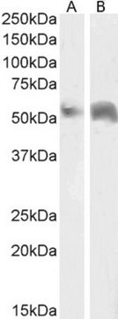
antigen ~62 kDa (also band at 46 kDa)

species reactivity

mouse

technique(s)

indirect immunofluorescence: 5-10 μg/mL using differentiated mouse NIH3T3-L1 cells
western blot (chemiluminescent): 2.5-5 μg/mL using whole extract of differentiated mouse NIH3T3-L1 cells

shipped in

dry ice

storage temp.

−20°C

target post-translational modification

unmodified

Gene Information

human ... PLIN(5346)
mouse ... Plin(103968)
rat ... Plin(25629)

General description

Perilipin is an intracellular neutral lipid storage droplet surface protein in white and brown fat adipocytes. It is also found in lower quantity coating droplets in steroidogenic-cells of the adrenal cortex, ovaries, and testicular Leydig cells, on the surface of smaller droplets containing cholesteryl esters.

Specificity

Rabbit polyclonal anti-Perilipin A/B antibody recognizes mouse Perilipin A/B by immunoblotting (46 kDa and ~62 kDa) and indirect immunofluorescence. Detection of the perilipin A and B bands by immunoblotting is specifically inhibited with the immunizing peptide.

Immunogen

synthetic peptide corresponding amino acid residues 6-17 of mouse and rat perilipin A/B with C-terminal added cysteine, conjugated to KLH. The corresponding human sequence differs by two residues.

Application

Anti-Perilipin A/B antibody produced in rabbit has been used in immunoblotting and immunofluorescence staining.
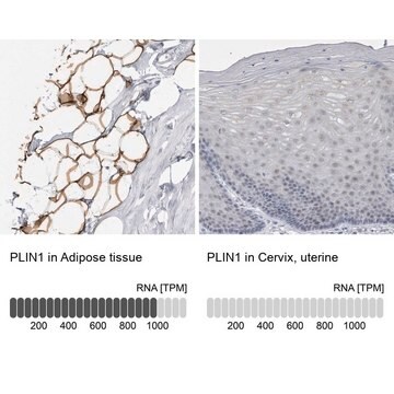
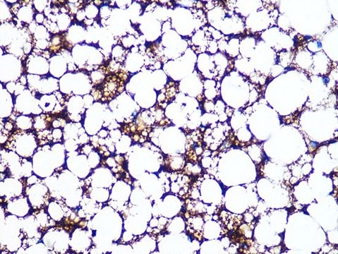
Rabbit polyclonal anti-Perilipin A/B antibody is used to tag Perilipin A/B for detection and quantitation by immunocytochemical and immunohistochemical (IHC) techniques. It is used as a probe to determine the presence and roles of Perilipin A/B in the regulation of triacylglycerol storage in adipocytes.

Biochem/physiol Actions

Perilipin is a gatekeeper protein that is involved in regulating triacylglycerol storage in adipocyte through the suppression of basal lipolysis apparently through protecting triacylglycerol against hydrolysis. Perilipin also enhances cAMP-dependent protein kinase (PKA)-stimulated lipolysis by hormone-sensitive lipase (HSL) and non-HSLs. Perilipin knockout mice exhibit reduced adipose tissue mass and resistance to diet induced obesity. Their lipid storage droplets are coated with adipose differentiation-related protein (ADRP, adipophilin), that is devoid of phosphorylation by PKA.

Physical form

Solution in 0.01 M phosphate buffered saline, pH 7.4, containing 15 mM sodium azide.

Disclaimer

Unless otherwise stated in our catalog or other company documentation accompanying the product(s), our products are intended for research use only and are not to be used for any other purpose, which includes but is not limited to, unauthorized commercial uses, in vitro diagnostic uses, ex vivo or in vivo therapeutic uses or any type of consumption or application to humans or animals.

Not finding the right product?  

Try our Product Selector Tool.

Storage Class Code

12 - Non Combustible Liquids

WGK

WGK 1

Flash Point(F)

Not applicable

Flash Point(C)

Not applicable

Personal Protective Equipment

dust mask type N95 (US), Eyeshields, Gloves

Certificates of Analysis (COA)

Search for Certificates of Analysis (COA) by entering the products Lot/Batch Number. Lot and Batch Numbers can be found on a product’s label following the words ‘Lot’ or ‘Batch’.

Already Own This Product?

Find documentation for the products that you have recently purchased in the Document Library.

Visit the Document Library

Takehiro Kasai et al.
The American journal of pathology, 187(12), 2627-2634 (2017-09-19)
Fatty degeneration of skeletal muscle leads to muscle weakness and loss of function. Preventing fatty degeneration in skeletal muscle is important, but no drug has been used clinically. In this study, we performed drug repositioning using human platelet-derived growth factor
Lijun Wang et al.
PLoS biology, 16(11), e2006522-e2006522 (2018-11-14)
During the aging process, bone marrow mesenchymal stem cells (BMSCs) exhibit declined osteogenesis accompanied by excess adipogenesis, which will lead to osteoporosis. Here, we report that the H3 lysine 36 trimethylation (H3K36me3), catalyzed by histone methyltransferase SET-domain-containing 2 (SETD2), regulates
Christian Elabd et al.
Nature communications, 5, 4082-4082 (2014-06-11)
The regenerative capacity of skeletal muscle declines with age. Previous studies suggest that this process can be reversed by exposure to young circulation; however, systemic age-specific factors responsible for this phenomenon are largely unknown. Here we report that oxytocin--a hormone
Reestablishment of energy balance in a male mouse model with POMC neuron deletion of BMPR1A
Townsend KL, et al.
Endocrinology, 158(12), 4233-4245 (2017)
The amino and carboxyl termini of perilipin a facilitate the storage of triacylglycerols
Garcia A, et al.
The Journal of Biological Chemistry, 279(9), 8409-8416 (2004)

Our team of scientists has experience in all areas of research including Life Science, Material Science, Chemical Synthesis, Chromatography, Analytical and many others.

Contact Technical Service