DUO92005
Duolink® In Situ PLA® Probe Anti-Rabbit MINUS
Synonym(s):
in situ Proximity Ligation Assay reagent, Protein Protein Interaction Assay reagent
About This Item
Recommended Products
biological source
donkey (polyclonal)
antibody form
affinity purified immunoglobulin (secondary antibody)
antibody product type
primary antibodies
product line
Duolink®
species reactivity
rabbit
technique(s)
immunofluorescence: suitable
proximity ligation assay: suitable
suitability
suitable for brightfield
suitable for fluorescence
shipped in
wet ice
storage temp.
2-8°C
Application
This product can be applied to both the Duolink® In Situ Fluorescence Protocol and the Duolink® In Situ Brightfield Protocol depending on the detection reagents used.
Visit our Duolink® PLA Resource Center for information on how to run a Duolink® experiment, applications, troubleshooting, and more.
To perform a complete Duolink® PLA in situ experiment you will need two primary antibodies (PLA, IHC, ICC or IF validated) that recognize two target epitopes. Other necessary reagents include a pair of PLA probes from different species (one PLUS and one MINUS), detection reagents, wash buffers, and mounting medium. Note that the primary antibodies must come from the same species as the Duolink® PLA probes. Analysis is carried out using standard immunofluorescence assay equipment.HRP is also available for brightfield detection.
PLA probe anti-Rabbit reacts with whole molecule rabbit IgG and the light chains of other rabbit immunoglobulin?s. The PLA probe anti-Rabbit has minimal cross reactivity with bovine, chicken, goat, guinea pig, Syrian hamster, horse, human, mouse, rat, and sheep serum proteins. A PLUS probe of a different species must be used simultaneously with this product. See our Product Selection Guide for more information.
Application Note
Two primary antibodies raised in different species are needed. Test your primary antibodies (IgG-class, mono- or polyclonal) in a standard immunofluorescence (IF), immunohistochemistry (IHC) or immunocytochemistry (ICC) assay to determine the optimal fixation, blocking, and titer conditions. Duolink® PLA in situ reagents are suitable for use on fixed cells, cytospin cells, cells grown on slide, formalin-fixed, paraffin embedded (FFPE), or tissue (fresh or frozen). No minimum number of cells is required.
Let us do the work for you, learn more about our Custom Service Program to accelerate your Duolink® projects
View full Duolink® product list
Features and Benefits
- No overexpression or genetic manipulation required
- High specificity (fewer false positives)
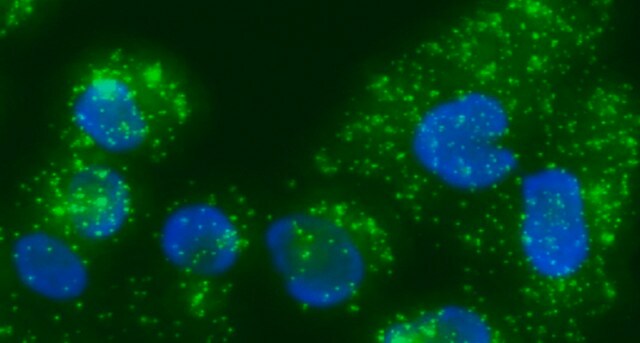
- Single molecule sensitivity due to rolling circle amplification
- Relative quantification possible
- No special equipment needed
- Quicker and simpler than FRET
- Increased accuracy compared to co-IP
- Publication-ready results
Components
- 5x PLA Probe Anti-Rabbit MINUS - Donkey anti-rabbit secondary antibody conjugated to oligonucleotide MINUS
- 1x Blocking Solution - Reagent for blocking of the sample
- 1x Antibody Diluent - For dilution of PLA probes and primary antibodies
Legal Information
Not finding the right product?
Try our Product Selector Tool.
Storage Class Code
10 - Combustible liquids
Certificates of Analysis (COA)
Search for Certificates of Analysis (COA) by entering the products Lot/Batch Number. Lot and Batch Numbers can be found on a product’s label following the words ‘Lot’ or ‘Batch’.
Already Own This Product?
Find documentation for the products that you have recently purchased in the Document Library.
Customers Also Viewed
Our team of scientists has experience in all areas of research including Life Science, Material Science, Chemical Synthesis, Chromatography, Analytical and many others.
Contact Technical Service


