Campylobacter Detection and Identification
Detection, identification, differentiation and cultivation of Campylobacter
The Campylobacter is one of the leading causes of human gastroenteritis. Common Campylobacter species C. jejuni, C. coli, and C. lari are responsible for most cases of campylobacteriosis.1,2 However, other species, like C. fetus, which causes spontaneous abortions, have also been associated with human illness.2–6
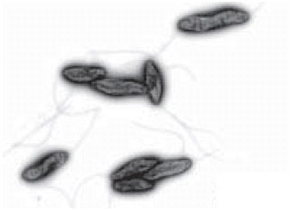
Campylobacter are Gram-negative, spiral-shaped, microaerophilic and motile bacteria with uni- or bi-polar flagella (Figures 1, 2 and 3).

Figure 1.Scanning electron microscope image: shows the characteristic spiral, or corkscrew, shape of C. jejuni cells. Photo by De Wood; digital colorization by Chris Pooley, USDA/ARS

Figure 2.Scanning electron micrograph of the single polar flagellum and corkscrew shape of C. jejuni. These morphologic characteristics contribute to the characteristic darting motility of C. jejuni in the viscous mucous layer of the intestinal lumen. Sean F. Altekruse National Cancer Institute, Rockville.

Figure 3.Electron microscope image of Campylobacters by Jochen Reetz, Bundesinstitut für Risikobewertung (BfR), Germany
The size of a cell is roughly 0.2–0.8 x 0.5–5 µm and, in culture, they can undergo morphological change from spiral to spherical. Most species are catalase- and oxidase-positive,7 with the exception of the catalase-negative C. sputorum, C. concisus, C. mucosalis and C. helveticus. The metabolism of Campylobacter is chemoorganotrophic, with amino acids and intermediates of the citric acid cycle serving as energy sources; typical carbohydrates cannot be used. Campylobacter reduce nitrate to nitrite, obtaining oxygen for their metabolism by this pathway. These distinctive metabolic reactions can be used for the differentiation and identification of Campylobacter species (Table 1). Commercially available tests are listed in Tables 2 and 3.

Table 1.Table of differentiating characteristics of Campylobacter species and subspecies
| Campylobacter test | Product No. |
|---|---|
| Catalase Test | 88597 |
| Hippurate Disks | 40405 |
| Hydrogen Sulfide Test Strips | 06728 |
| Indoxyl Strips | 04739 |
| Nitrate Reagent A | 38497 |
| Nitrate Reagent B | 39441 |
| Nitrate Reagent Disks | 08086 |
| Oxidase Test | 70439 |
| Oxidase Strips | 40560 |
| Oxidase Reagent acc. Gaby-Hadley A | 07345 |
| Oxidase Reagent acc. Gaby-Hadley B | 07817 |
| Oxidase Reagent acc. Gordon-McLeod | 18502 |
Campylobacter are generally fastidious microorganisms and grow only on complex media that have been amended with diverse essential amino acids and supplements, such as pyruvate, a-ketoglutarate, hemin, formate and other essential ions. For selective isolation of Campylobacter, the growth media can be supplemented with antibiotics like cefoperazone, vancomycin, trimethoprim, amphotericin, cycloheximide, rifampicin, cefsulodin and polymyxin B sulfate. We offer a broad range of specific agars and broths for the detection, identification, differentiation, enumeration and cultivation of Campylobacter (Table 4).
| Nonselective media | Product No. |
|---|---|
| Tryptic Soy Broth, Vegitone | 41298 |
| Columbia Agar | 27688 |
| Tryptic Soy Agar, Vegitone | 14432 |
| Tryptic Soy Broth | 22092 |
| CASO Agar | 22095 |
| Tryptic Soy Broth No. 2 | 51228 |
| Tryptic Soy Agar | 22091 |
| Tryptic Soy Agar Plates (Diameter 55 mm) | 57994 |
| CASO Broth | 22098 |
| Nonselective nedia with differential system | |
| Blood Agar Base No. 2 | |
| Hippurate Broth | 53275 |
| OF Test Nutrient Agar | 75315 |
| Selective media with differential system | |
| MacConkey Agar No. 1 | |
| Campylobacter selective media | |
| Bolton Broth Base | |
| Karmali Campylobacter Agar (Base) | 17152 |
| Brucella Broth Base | B3051 |
| Campylobacter Selective Agar (Base) | 21378 |
| Park and Sanders Enrichment Broth (Base) | 17189 |
| Blood-free Campylobacter Selective Agar Base | B2426 |
More details about our products for analytical microbiology.
| Kingdom: | Bacteria |
| Phylum: | Proteobacteria |
| Class: | Epsilon Proteobacteria |
| Order: | Campylobacterales |
| Family: | Campylobacteraceae |
| Genus: | Campylobacter |
Network error: Failed to fetch
References
계속 읽으시려면 로그인하거나 계정을 생성하세요.
계정이 없으십니까?