
HPA019852
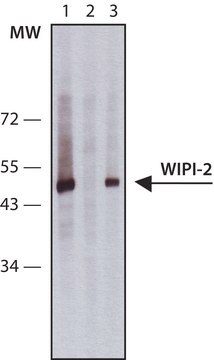
Anti-WIPI2 antibody produced in rabbit
Prestige Antibodies® Powered by Atlas Antibodies, affinity isolated antibody, buffered aqueous glycerol solution, Ab1
동의어(들):
Anti-WD repeat domain phosphoinositide-interacting protein 2, Anti-WIPI-2, Anti-WIPI49-like protein 2
로그인조직 및 계약 가격 보기
모든 사진(5)
About This Item
추천 제품


생물학적 소스
rabbit
Quality Level
결합
unconjugated
항체 형태
affinity isolated antibody
항체 생산 유형
primary antibodies
클론
polyclonal
제품 라인
Prestige Antibodies® Powered by Atlas Antibodies
양식
buffered aqueous glycerol solution
종 반응성
rat, mouse, human
기술
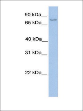
immunoblotting: 0.04-0.4 μg/mL
immunofluorescence: 0.25-2 μg/mL
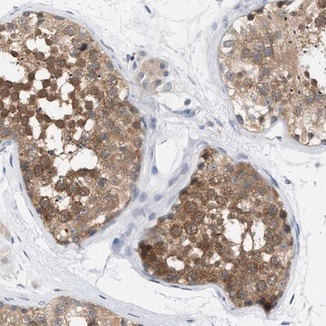
immunohistochemistry: 1:50-1:200
면역원 서열
ECALMKQHRLDGSLETTNEILDSASHDCPLVTQTYGAAAGKGTYVPSSPTRLAYTDDLGAVGGACLEDEASALRLDEDSEHPPMILRTD
UniProt 수납 번호
배송 상태
wet ice
저장 온도
−20°C
타겟 번역 후 변형
unmodified
유전자 정보
human ... WIPI2(26100)
일반 설명
WIPI2 (WD repeat domain, phosphoinositide interacting 2) is a WD-repeat PtdIns(3)P (phosphatidylinositol 3-phosphate) effector protein expressed in the endoplasmic reticulum. It is expressed in different cell lines. WIPI2 exists in five isoforms (a, b, c, d, and e).
면역원
WD repeat domain phosphoinositide-interacting protein 2 recombinant protein epitope signature tag (PrEST)
애플리케이션
All Prestige Antibodies Powered by Atlas Antibodies are developed and validated by the Human Protein Atlas (HPA) project and as a result, are supported by the most extensive characterization in the industry.
The Human Protein Atlas project can be subdivided into three efforts: Human Tissue Atlas, Cancer Atlas, and Human Cell Atlas. The antibodies that have been generated in support of the Tissue and Cancer Atlas projects have been tested by immunohistochemistry against hundreds of normal and disease tissues and through the recent efforts of the Human Cell Atlas project, many have been characterized by immunofluorescence to map the human proteome not only at the tissue level but now at the subcellular level. These images and the collection of this vast data set can be viewed on the Human Protein Atlas (HPA) site by clicking on the Image Gallery link. We also provide Prestige Antibodies® protocols and other useful information.
The Human Protein Atlas project can be subdivided into three efforts: Human Tissue Atlas, Cancer Atlas, and Human Cell Atlas. The antibodies that have been generated in support of the Tissue and Cancer Atlas projects have been tested by immunohistochemistry against hundreds of normal and disease tissues and through the recent efforts of the Human Cell Atlas project, many have been characterized by immunofluorescence to map the human proteome not only at the tissue level but now at the subcellular level. These images and the collection of this vast data set can be viewed on the Human Protein Atlas (HPA) site by clicking on the Image Gallery link. We also provide Prestige Antibodies® protocols and other useful information.
생화학적/생리학적 작용
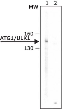
WIPI2 (WD repeat domain, phosphoinositide interacting 2) is involved in the regulation of starvation-induced autophagy. It directly interacts to the Atg12-5-16L1 complex upstream of Atg16L1. During starvation-induced autophagy, this interaction further facilitates PtdIns(3)P-dependent recruitment of Atg12-5-16L1 to the omegasome and phagophore. It also plays an essential role in the innate immune response to Salmonella Typhimurium infection by initiating several responses such as the introduction of Atg16L1, initiation of LC3 lipid conjugation, autophagosomal membrane formation, and blocking of bacterial proliferation.
특징 및 장점
Prestige Antibodies® are highly characterized and extensively validated antibodies with the added benefit of all available characterization data for each target being accessible via the Human Protein Atlas portal linked just below the product name at the top of this page. The uniqueness and low cross-reactivity of the Prestige Antibodies® to other proteins are due to a thorough selection of antigen regions, affinity purification, and stringent selection. Prestige antigen controls are available for every corresponding Prestige Antibody and can be found in the linkage section.
Every Prestige Antibody is tested in the following ways:
Every Prestige Antibody is tested in the following ways:
- IHC tissue array of 44 normal human tissues and 20 of the most common cancer type tissues.
- Protein array of 364 human recombinant protein fragments.
결합
Corresponding Antigen APREST75037
물리적 형태
Solution in phosphate-buffered saline, pH 7.2, containing 40% glycerol and 0.02% sodium azide
법적 정보
Prestige Antibodies is a registered trademark of Merck KGaA, Darmstadt, Germany
면책조항
Unless otherwise stated in our catalog or other company documentation accompanying the product(s), our products are intended for research use only and are not to be used for any other purpose, which includes but is not limited to, unauthorized commercial uses, in vitro diagnostic uses, ex vivo or in vivo therapeutic uses or any type of consumption or application to humans or animals.
적합한 제품을 찾을 수 없으신가요?
당사의 제품 선택기 도구.을(를) 시도해 보세요.
Storage Class Code
10 - Combustible liquids
WGK
WGK 1
Flash Point (°F)
Not applicable
Flash Point (°C)
Not applicable
가장 최신 버전 중 하나를 선택하세요:
Hannah C Dooley et al.
Molecular cell, 55(2), 238-252 (2014-06-24)
Mammalian cell homeostasis during starvation depends on initiation of autophagy by endoplasmic reticulum-localized phosphatidylinositol 3-phosphate (PtdIns(3)P) synthesis. Formation of double-membrane autophagosomes that engulf cytosolic components requires the LC3-conjugating Atg12-5-16L1 complex. The molecular mechanisms of Atg12-5-16L1 recruitment and significance of PtdIns(3)P
Megha Bansal et al.
The Journal of biological chemistry, 293(1), 132-147 (2017-11-15)
Autophagy is a quality-control mechanism that helps to maintain cellular homeostasis by removing damaged proteins and organelles through lysosomal degradation. During autophagy, signaling events lead to the formation of a cup-shaped structure called the phagophore that matures into the autophagosome.
Maria Giovanna De Leo et al.
Autophagy, 17(11), 3644-3670 (2021-03-10)
Autophagosome formation requires PROPPIN/WIPI proteins and monophosphorylated phosphoinositides, such as phosphatidylinositol-3-phosphate (PtdIns3P) or PtdIns5P. This process occurs in association with mammalian endosomes, where the PROPPIN WIPI1 has additional, undefined roles in vesicular traffic. To explore whether these functions are interconnected
자사의 과학자팀은 생명 과학, 재료 과학, 화학 합성, 크로마토그래피, 분석 및 기타 많은 영역을 포함한 모든 과학 분야에 경험이 있습니다..
고객지원팀으로 연락바랍니다.