추천 제품
생물학적 소스
mouse
Quality Level
결합
unconjugated
항체 형태
ascites fluid
항체 생산 유형
primary antibodies
클론
CY-90, monoclonal
포함
15 mM sodium azide
종 반응성
wide range
기술
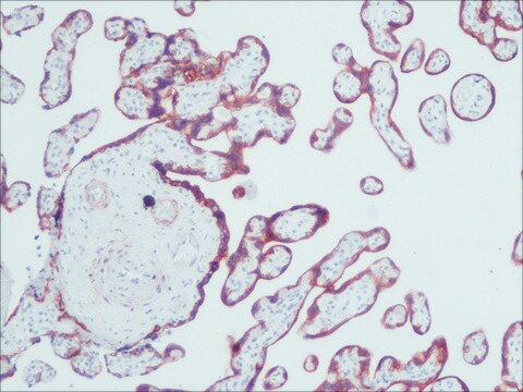
indirect immunofluorescence: 1:800 using formalin-fixed, paraffin-embedded sections of human tissue
western blot: suitable
동형
IgG1
배송 상태
dry ice
저장 온도
−20°C
타겟 번역 후 변형
unmodified
유사한 제품을 찾으십니까? 방문 제품 비교 안내
일반 설명
Cytokeratin 18 (45 kDa) is the primary type I keratin expressed in simple epithelial cells. Epithelial cells and their derivatives characteristically contain intermediate filaments (IFs) composed of about 20 related polypeptides with molecular weights between 40,000-69,000. Each epithelial tissue has a specific and stable pattern of expression of some of these cytokeratin subunits. Epithelium derived tumors maintain the expression of the cytokeratins found in the normal tissue of origin. Therefore, carcinomas can be identified and classified by immunocytochemical staining with antibodies that react specifically with cytokeratins.
Monoclonal Anti-Cytokeratin Peptide 18 (mouse IgG1 isotype) is derived from the hybridoma produced by the fusion of mouse myeloma cells and splenocytes from an immunized mouse. Cytokeratin 18 (CK18)/keratin 18 is a structural protein and is present in single-layer epithelia. It is located on human chromosome 12q13.
특이성
The antibody reacts specifically with a wide variety of simple epithelia (e.g. intestine, respiratory and urinary systems, liver, and glandular epithelia). It does not react with stratified squamous epithelia (e.g. esophagus or epidermis) or with non-epithelial cells.
면역원
human epidermal carcinoma A-431 and MCF-7 human breast cancer cell lines.
애플리케이션
Monoclonal Anti-Cytokeratin Peptide 18 antibody produced in mouse has been used in:
- immunofluorescence
- fluorescence microscopy
- confocal fluorescence microscopy
- immunolabelling
- histoblots
생화학적/생리학적 작용
Keratins help to maintain structural integrity. They also control Fas?mediated apoptosis and regulate cell size and protein synthesis. Cytokeratin 18 (CK18) plays a major role in the development of lung cancer. It acts as a therapeutic target for non-small cell lung cancer (NSCLC). CK-18 is considered as a promising plasma biomarker to identify patients with HELLP (hemolysis, elevated liver enzymes, low platelets) syndrome.
면책조항
Unless otherwise stated in our catalog or other company documentation accompanying the product(s), our products are intended for research use only and are not to be used for any other purpose, which includes but is not limited to, unauthorized commercial uses, in vitro diagnostic uses, ex vivo or in vivo therapeutic uses or any type of consumption or application to humans or animals.
적합한 제품을 찾을 수 없으신가요?
당사의 제품 선택기 도구.을(를) 시도해 보세요.
Storage Class Code
10 - Combustible liquids
WGK
WGK 3
Flash Point (°F)
Not applicable
Flash Point (°C)
Not applicable
시험 성적서(COA)
제품의 로트/배치 번호를 입력하여 시험 성적서(COA)을 검색하십시오. 로트 및 배치 번호는 제품 라벨에 있는 ‘로트’ 또는 ‘배치’라는 용어 뒤에서 찾을 수 있습니다.
H Inada et al.
The Journal of cell biology, 155(3), 415-426 (2001-10-31)
Keratin 8 and 18 (K8/18) are the major components of intermediate filament (IF) proteins of simple or single-layered epithelia. Recent data show that normal and malignant epithelial cells deficient in K8/18 are nearly 100 times more sensitive to tumor necrosis
Immuno-electron microscopic localisation of caveolin 1 in human placenta
Byrne S, et al.
Immunobiology, 212(1), 39-46 (2007)
Matthew J Lawson et al.
Royal Society open science, 8(11), 211067-211067 (2021-11-06)
Micro-computed tomography (µCT) provides non-destructive three-dimensional (3D) imaging of soft tissue microstructures. Specific features in µCT images can be identified using correlated two-dimensional (2D) histology images allowing manual segmentation. However, this is very time-consuming and requires specialist knowledge of the
Detection of PrPSc in rectal biopsy and necropsy samples from sheep with experimental scrapie
Espenes A, et al.
Journal of Comparative Pathology, 134(2-3), 115-125 (2006)
Thomas Brabletz et al.
Cancer research, 64(19), 6973-6977 (2004-10-07)
The homeobox transcription factor Cdx2 specifies intestinal development and homeostasis and is considered a tumor suppressor in colorectal carcinogenesis. However, Cdx2 mutations are rarely found. Invasion of colorectal cancer is characterized by a transient loss of differentiation and nuclear accumulation
자사의 과학자팀은 생명 과학, 재료 과학, 화학 합성, 크로마토그래피, 분석 및 기타 많은 영역을 포함한 모든 과학 분야에 경험이 있습니다..
고객지원팀으로 연락바랍니다.


