추천 제품
생물학적 소스
rabbit
Quality Level
항체 형태
purified antibody
항체 생산 유형
primary antibodies
클론
Vli-55, monoclonal
종 반응성
rat, mouse, human, porcine
기술
immunohistochemistry: suitable
western blot: suitable
NCBI 수납 번호
UniProt 수납 번호
배송 상태
wet ice
타겟 번역 후 변형
unmodified
유전자 정보
mouse ... Cthrc1(68588)
일반 설명
Collagen triple helix repeat-containing protein 1 (UniProt Q96CG8; also known as Protein NMTC1) is encoded by the CTHRC1 gene (ORF UNQ762/PRO1550; Gene ID 115908) in human. CTHRC1 is a secreted glycoprotein with 12 repeats of the Gly-X-Y motif (a.a. 58-93 based on the pro-form seqeunce) that is reported to increase cell motility and promote cell migration by limiting the deposition of collagen matrix. CTHRC1 is normally not expressed in blood vessels, but is induced in adventitial cells in remodeling arteries, as well as in dermal fibroblasts during wound healing. Upregulatetd CTHRC1 level is also observed in many human solid tumors, including non-small-cell lung cancer (NSCLC), gastric cancer, hepatocellular cancer, breast cancer, pancreatic cancer, and colorectal cancer (CRC). CTHRC1 is initially produced with a signal peptide sequence (a.a. 1-30), the removal of which yields the 213-amino acid (a.a. 31-243) mature protein.
특이성
Clone Vli-55 stained the midbrain region of wild-type, but not Cthrc1-null mice (Stohn, J.P., et al. (2012). PLoS One. 7(10):e47142). Expected to react with all three spliced isoforms of human CTHRC1 reported by UniProt (Q96CG8).
면역원
Epitope: Near C-terminus.
Linear peptide corresponding to a C-terminal sequence of human CTHRC1.
애플리케이션
Anti-CTHRC1 Antibody, clone Vli-55 is an antibody against CTHRC1 for use in Western Blotting, Immunohistochemistry.
Immunohistochemistry Analysis: Clone Vli-55 hybridoma culture supernatant (1:250 dilution) immunostained the paraventricular nucleus of the hypothalamus in mouse brain tissue sections (Courtesy of Dr. Volkhard Lindner, Tufts University School of Medicine, ME, USA).
Western Blotting Analysis: Representative lots detected exogenously expressed human and rat CTHRC1 in lysates from transfected cells (Duarte, C.W., et al. (2014). PLoS One.;9(6):e100449; Stohn, J.P., et al. (2012). PLoS One. 7(10):e47142).
Western Blotting Analysis: A representative lot detected the overexpressed Cthrc1 in plasma samples from Cthrc1 trangenic mice, as well as immunoprecipitated CTHRC1 from normal human plasma samples (Stohn, J.P., et al. (2012). PLoS One. 7(10):e47142).
Immunohistochemistry Analysis: A representative lot detected increased Cthrc1 expression in interstitial cells and activated fibroblasts of various remodeling tissues from wild-type, but not Cthrc1-null mice following angiotensin II infusion by immunohistochemistry staining of paraformaldehyde-fixed, paraffin-embedded sections (Duarte, C.W., et al. (2014). PLoS One.;9(6):e100449).
Immunohistochemistry Analysis: Representative lots immunostained Cthrc1-positive cells in paraformaldehyde-fixed, paraffin-embedded human, pig, and rat pituitary gland tissue sections (Duarte, C.W., et al. (2014). PLoS One.;9(6):e100449; Stohn, J.P., et al. (2012). PLoS One. 7(10):e47142).
Immunohistochemistry Analysis: A representative lot immunostained adventitial cells of remodeling renal artery, dermal cells in skin wounds, embryonic cartilage, as well as midbrain of wild-type, but not Cthrc1-null mice, using paraformaldehyde-fixed, paraffin-embedded tissue sections (Stohn, J.P., et al. (2012). PLoS One. 7(10):e47142).
Immunohistochemistry Analysis: A representative lot localized Cthrc1 immunoreactivity in various regions of paraformaldehyde-fixed, paraffin-embedded mouse and pig brain sections (Stohn, J.P., et al. (2012). PLoS One. 7(10):e47142).
Research Category
Cell Structure
Cell Structure
Research Sub Category
Adhesion (CAMs)
Adhesion (CAMs)
품질
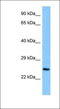
Evaluated by Western Blotting in human brain tissue lysate.
Western Blotting Analysis: 10 µg/mL of this antibody detected CTHRC1 in 10 µg of human brain tissue lysate.
Western Blotting Analysis: 10 µg/mL of this antibody detected CTHRC1 in 10 µg of human brain tissue lysate.
표적 설명
~25 kDa observed. 23.08 kDa (isoform 1), 25.16 kDa (isoform 2), 24.77 kDa (isoform 3) calculated.
물리적 형태
Format: Purified
Protein G Purified
Purified rabbit monoclonal antibody in buffer containing 0.1 M Tris-Glycine (pH 7.4), 150 mM NaCl with 0.05% sodium azide.
저장 및 안정성
Stable for 1 year at 2-8°C from date of receipt.
기타 정보
Concentration: Please refer to lot specific datasheet.
면책조항
Unless otherwise stated in our catalog or other company documentation accompanying the product(s), our products are intended for research use only and are not to be used for any other purpose, which includes but is not limited to, unauthorized commercial uses, in vitro diagnostic uses, ex vivo or in vivo therapeutic uses or any type of consumption or application to humans or animals.
Not finding the right product?
Try our 제품 선택기 도구.
Storage Class Code
12 - Non Combustible Liquids
WGK
WGK 1
Flash Point (°F)
Not applicable
Flash Point (°C)
Not applicable
시험 성적서(COA)
제품의 로트/배치 번호를 입력하여 시험 성적서(COA)을 검색하십시오. 로트 및 배치 번호는 제품 라벨에 있는 ‘로트’ 또는 ‘배치’라는 용어 뒤에서 찾을 수 있습니다.
Huixin Li et al.
Oncology letters, 22(6), 814-814 (2021-10-22)
Cancer-associated fibroblasts (CAFs) are continuously activated and are one of the most important cellular components of the tumor matrix. The role of CAFs in the tumor microenvironment has been widely recognized. However, the underlying molecular mechanism by which CAFs promote
자사의 과학자팀은 생명 과학, 재료 과학, 화학 합성, 크로마토그래피, 분석 및 기타 많은 영역을 포함한 모든 과학 분야에 경험이 있습니다..
고객지원팀으로 연락바랍니다.