추천 제품
생물학적 소스
mouse
Quality Level
항체 형태
purified immunoglobulin
항체 생산 유형
primary antibodies
클론
N106/36, monoclonal
종 반응성
rat
기술
immunohistochemistry: suitable
동형
IgG2aκ
NCBI 수납 번호
UniProt 수납 번호
배송 상태
wet ice
타겟 번역 후 변형
unmodified
유전자 정보
rat ... Ank3(361833)
일반 설명
Ankyrins are a family of spectrin-binding proteins that couple the spectrin/actin cytoskeletal network to the cytoplasmic domains of integral membrane proteins which include channels such as the anion exchanger, voltage-dependent sodium channel, and the Na/K ATPase, as well as neuronal cell adhesion molecules related to L1. In the nervous system, three different ankyrins are known to exist; ankyrinB, ankyrinG, and ankyrinR. All three ankyrins contain an N-terminal membrane binding domain, an internal domain capable of binding spectrin, and a C-terminal regulatory domain which is the target of alternative splicing. AnkyrinB, the predominant neuronal ankyrin includes two isoforms of ~220 kDa and ~440 kDa. The ~220 kDa alternatively spliced isoform is the major form present in the adult brain, while the ~440 kDa form is preferentially expressed in the developing neonatal brain. Three forms of ankyrinG (~190 kDa, ~270 kDa, ~480 kDa) are localized in axonal initial segments and the nodes of Ranvier of myelinated axons where fluxes of action potential occur and ion channels are enriched. Ankyrins comprise ~0.5%-1% of the total membrane protein in the adult vertebrate brain.
특이성
Other homologies: Human (92% sequence homology), and Mouse (87% sequence homology).
면역원
Recombinant protein corresponding to human Ankyrin-G.
애플리케이션
Anti-Ankyrin-G Antibody, clone N106/36 is a highly specific mouse monoclonal antibody & that targets Ankyrin & has been tested in IHC.
Immunohistochemistry Analysis: A 1:2,000 dilution from a representative lot detected Ankyrin-G in rat cerebral cortex tissue.
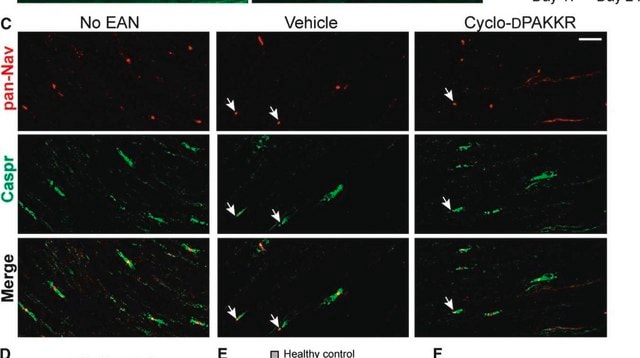
Immunohistochemistry Analysis: A representative lot detected Ankyrin-G in rat optic nerve tissue.
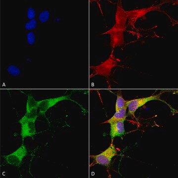
Immunofluorescence Analysis: A representative lot detected Ankyrin-G in adult rat cortex tissue.
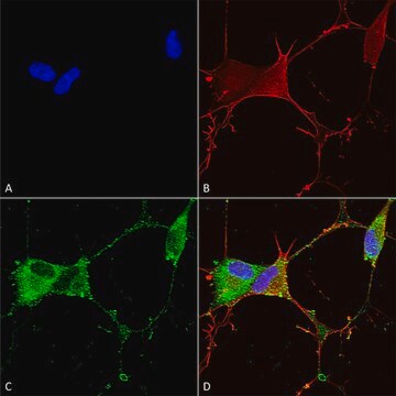
Immunofluorescence Analysis: A representative lot from an independent laboratory detected Ankyrin-G in a rat model of TLE mEC layer II neurons (Hargus, N. J., et al. (2011). Neurobiol Dis. 41(2):361-376.)
Research Category
Neuroscience
Neuroscience
Research Sub Category
Developmental Neuroscience
Developmental Neuroscience
품질
Evaluated by Immunohistochemistry in rat hippocampus tissue.
Immunohistochemistry Analysis: A 1:500 dilution from a representative lot detected Ankyrin-G in rat hippocampus tissue.
Immunohistochemistry Analysis: A 1:500 dilution from a representative lot detected Ankyrin-G in rat hippocampus tissue.
표적 설명
480 kDa calculated
물리적 형태
Format: Purified
Protein G Purified
Purified mouse monoclonal IgG2a κ in 0.1 M Tris-Glycine (pH 7.4), 150 mM NaCl with 0.05% sodium azide.
저장 및 안정성
Stable for 1 year at 2-8°C from date of receipt.
분석 메모
Control
Rat hippocampus tissue
Rat hippocampus tissue
기타 정보
Concentration: Please refer to the Certificate of Analysis for the lot-specific concentration.
면책조항
Unless otherwise stated in our catalog or other company documentation accompanying the product(s), our products are intended for research use only and are not to be used for any other purpose, which includes but is not limited to, unauthorized commercial uses, in vitro diagnostic uses, ex vivo or in vivo therapeutic uses or any type of consumption or application to humans or animals.
Not finding the right product?
Try our 제품 선택기 도구.
Storage Class Code
12 - Non Combustible Liquids
WGK
WGK 1
Flash Point (°F)
Not applicable
Flash Point (°C)
Not applicable
시험 성적서(COA)
제품의 로트/배치 번호를 입력하여 시험 성적서(COA)을 검색하십시오. 로트 및 배치 번호는 제품 라벨에 있는 ‘로트’ 또는 ‘배치’라는 용어 뒤에서 찾을 수 있습니다.
Zhuang Xu et al.
iScience, 27(3), 109264-109264 (2024-03-07)
The axon initial segment (AIS) is located at the proximal axon demarcating the boundary between axonal and somatodendritic compartments. The AIS facilitates the generation of action potentials and maintenance of neuronal polarity. In this study, we show that the location
Yanxia Ding et al.
Neuroreport, 29(18), 1537-1543 (2018-10-16)
Recent studies have indicated that the structure of the axon initial segment (AIS) of neurons is highly plastic in response to changes in neuronal activity. Whether an age-related enhancement of neuronal responses in the visual cortex is coupled with plasticity
Anja Konietzny et al.
Journal of cell science, 137(8) (2024-03-25)
In neurons, the microtubule (MT) cytoskeleton forms the basis for long-distance protein transport from the cell body into and out of dendrites and axons. To maintain neuronal polarity, the axon initial segment (AIS) serves as a physical barrier, separating the
Miguel A Marin et al.
The Journal of neuroscience : the official journal of the Society for Neuroscience, 43(48), 8126-8139 (2023-10-12)
Subcortical white matter stroke (WMS) is a progressive disorder which is demarcated by the formation of small ischemic lesions along white matter tracts in the CNS. As lesions accumulate, patients begin to experience severe motor and cognitive decline. Despite its
Tuancheng Feng et al.
Acta neuropathologica communications, 10(1), 33-33 (2022-03-16)
TMEM106B, a type II lysosomal transmembrane protein, has recently been associated with brain aging, hypomyelinating leukodystrophy, frontotemporal lobar degeneration (FTLD) and several other brain disorders. TMEM106B is critical for proper lysosomal function and TMEM106B deficiency leads to myelination defects, FTLD
자사의 과학자팀은 생명 과학, 재료 과학, 화학 합성, 크로마토그래피, 분석 및 기타 많은 영역을 포함한 모든 과학 분야에 경험이 있습니다..
고객지원팀으로 연락바랍니다.