
추천 제품


생물학적 소스
mouse
Quality Level
항체 형태
purified antibody
항체 생산 유형
primary antibodies
클론
1C4, monoclonal
종 반응성
rat, mouse, human
기술
immunocytochemistry: suitable
immunoprecipitation (IP): suitable
western blot: suitable
동형
IgG1κ
NCBI 수납 번호
UniProt 수납 번호
배송 상태
ambient
타겟 번역 후 변형
unmodified
유전자 정보
human ... NF2(4771)
일반 설명
Merlin (UniProt P35240; also known as Moesin-ezrin-radixin-like protein, Neurofibromin-2, Schwannomerlin, Schwannomin) is encoded by the NF2 (also known as SCH) gene (Gene ID 4771) in human. Originally identified as a tumor suppressor gene in the familial Neurofibromatosis type 2 (NF2) cancer patients, NF2 encodes the FERM (Ezrin, Radixin, Moesin) domain-containing merlin that plays a negative regulating role against cell growth and proliferation by functioning as an upstream activator of the Hippo pathway. Merlin directly activates Mst1/2 and recruits Lats1/2 to the plasma membrane for phosphorylation by Mst1/2. The phosphorylated Lats1/2 in turn inhibits YAP/TAZ transcription activity by phosphorylating YAP/TAZ and causes its cytoplasmic sequestration. Merlin also translocates to the nucleus, where it inhibits E3 ubiquitin ligase CRL4(DCAF1), thereby preventing it from ubiquitinating and downregulating Lats1/2. In its inactive state, the C-terminal tail domain (CTD; a.a. 507-595) of merlin is bound intramolecularly to its FERM domain (a.a. 19-313). Angiomotin (AMOT) induces merlin activation by binding to an elongated helical fragment within merlin CTD and subsequently releases the auto-inhibited conformation of Merlin. Merlin CTD can also undergo phosphorylation on Ser518 by p21-activated kinase (PAK) or cyclic AMP-dependent protein kinase (PKA), resulting in a weakened AMOT binding and a stabilization of merlin in its auto-inhibited state.
특이성
Clone 1C4 detected merlin (NF2), but not any of the three ERM proteins, ezrin, radixin, and moesin (Gonzalez-Agosti, C., et al. (1996). Oncogene. 13(6):1239-1247). Clone 1C4 targeted epitope is present in human merlin (NF2) spliced isoforms 1 to 6, but not 7, 9, or 10 as reported by UniProt (P35240). Reactivity toward human spliced isoform 8 is possible, but has not been confirmed.
면역원
Recombinant protein corresponding to the C-terminal half of human merlin (NF2) isoform II (UniProt spliced isoform 3; P35240-3).
애플리케이션
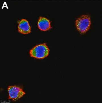
Anti-Merlin (NF2), clone 1C4, Cat. No. MABN1786, is a highly specific mouse monoclonal antibody that targets Merlin (NF2) and has been tested in Immunocytochemistry, Immunoprecipitation, and Western Blotting.
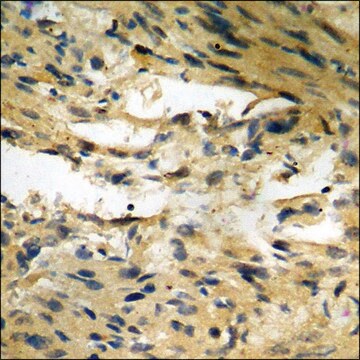
Immunocytochemistry Analysis: A representative lot immunostained % paraformaldehyde-fixed, 0.2% Triton X-100-permeabilized wild-type, but not Nf2-knockout, mouse liver-derived epithelial cells (LDCs) by fluorescent immunocytochemistry. EGF-induced EGFR internalization was observed among Nf2-/-, but not Nf2+/+, LDCs (Curto, M., et al. (2007). J. Cell Biol. 177(5):893-903).
Immunocytochemistry Analysis: A representative lot detected merlin cellular distribution and HEI10 co-localization in a cell cycle-dependent manner by fluorescent immunocytochemistry staining of 3.5% paraformaldehyde-fixed U2OS human osteosarcoma cells (Grönholm, M., et al. (2006). Oncogene. 25(32):4389-4398).
Immunocytochemistry Analysis: A representative lot detected merlin cellular localization by fluorescent immunocytochemistry staining of 3.5% paraformaldehyde-fixed embryonic E16 rat neurons. Merlin was seen co-localized with RI to the cell body and extensions in a punctate pattern (Grönholm, M., et al. (2003). J. Biol. Chem. 278(42):41167-41172).
Immunocytochemistry Analysis: A representative lot detected merlin cellular localization by fluorescent immunocytochemistry staining of 4% paraformaldehyde-fixed, 0.1 NP-40-permeabilized human fibroblasts and primary meningioma cells. Merlin co-localized with F-actin at the leading and ruffling edges, but not at the stress fiber. No merlin co-localization with ezrin or moesin was observed (Gonzalez-Agosti, C., et al. (1996). Oncogene. 13(6):1239-1247).
Immunoprecipitation Analysis: A representative lot co-immunoprecipitated N-WASP with merlin from HEK293T cell lysate (Manchanda, N., et al. (2005). J. Biol. Chem. 280(13):12517-12522).
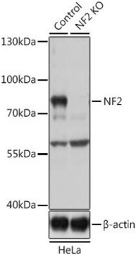
Western Blotting Analysis: A representative lot detected the presence of wild-type, but not L64P, merlin in the EGFR immunoprecipitates from Nf2-/- MEFs infected by adenovirus to express either wild-type or L64P merlin (Curto, M., et al. (2007). J. Cell Biol. 177(5):893-903).
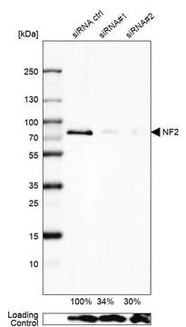
Western Blotting Analysis: A representative lot detected a reduced merlin level in EGFR immunoprecipitate from mouse liver-derived epithelial cells (LDCs) upon shRNA-mediated NHE-RF1, but not NHE-RF2 knockdown. Merlin association with Ezrin was not affected by NHE-RF1 knockdown (Curto, M., et al. (2007). J. Cell Biol. 177(5):893-903).
Western Blotting Analysis: A representative lot detected a ~72 kDa merlin band in rat newborn fibroblast (RNF) and S-16 rat Schwann cell lysates, as well as a ~66 kDa merlin band in murine NIH/3T3 and human MRC-5 fetal fibroblast lystes without cross-reactivity toward the three ERM protein family members, ezrin, radixin, and moesin (Gonzalez-Agosti, C., et al. (1996). Oncogene. 13(6):1239-1247).
Immunocytochemistry Analysis: A representative lot detected merlin cellular distribution and HEI10 co-localization in a cell cycle-dependent manner by fluorescent immunocytochemistry staining of 3.5% paraformaldehyde-fixed U2OS human osteosarcoma cells (Grönholm, M., et al. (2006). Oncogene. 25(32):4389-4398).
Immunocytochemistry Analysis: A representative lot detected merlin cellular localization by fluorescent immunocytochemistry staining of 3.5% paraformaldehyde-fixed embryonic E16 rat neurons. Merlin was seen co-localized with RI to the cell body and extensions in a punctate pattern (Grönholm, M., et al. (2003). J. Biol. Chem. 278(42):41167-41172).
Immunocytochemistry Analysis: A representative lot detected merlin cellular localization by fluorescent immunocytochemistry staining of 4% paraformaldehyde-fixed, 0.1 NP-40-permeabilized human fibroblasts and primary meningioma cells. Merlin co-localized with F-actin at the leading and ruffling edges, but not at the stress fiber. No merlin co-localization with ezrin or moesin was observed (Gonzalez-Agosti, C., et al. (1996). Oncogene. 13(6):1239-1247).
Immunoprecipitation Analysis: A representative lot co-immunoprecipitated N-WASP with merlin from HEK293T cell lysate (Manchanda, N., et al. (2005). J. Biol. Chem. 280(13):12517-12522).
Western Blotting Analysis: A representative lot detected the presence of wild-type, but not L64P, merlin in the EGFR immunoprecipitates from Nf2-/- MEFs infected by adenovirus to express either wild-type or L64P merlin (Curto, M., et al. (2007). J. Cell Biol. 177(5):893-903).
Western Blotting Analysis: A representative lot detected a reduced merlin level in EGFR immunoprecipitate from mouse liver-derived epithelial cells (LDCs) upon shRNA-mediated NHE-RF1, but not NHE-RF2 knockdown. Merlin association with Ezrin was not affected by NHE-RF1 knockdown (Curto, M., et al. (2007). J. Cell Biol. 177(5):893-903).
Western Blotting Analysis: A representative lot detected a ~72 kDa merlin band in rat newborn fibroblast (RNF) and S-16 rat Schwann cell lysates, as well as a ~66 kDa merlin band in murine NIH/3T3 and human MRC-5 fetal fibroblast lystes without cross-reactivity toward the three ERM protein family members, ezrin, radixin, and moesin (Gonzalez-Agosti, C., et al. (1996). Oncogene. 13(6):1239-1247).
Research Category
Neuroscience
Neuroscience
품질
Evaluated by Western Blotting in HEK293 cell lysate.
Western Blotting Analysis: 4 µg/mL of this antibody detected Merlin (NF2) in 10 µg of HEK293 cell lysate.
Western Blotting Analysis: 4 µg/mL of this antibody detected Merlin (NF2) in 10 µg of HEK293 cell lysate.
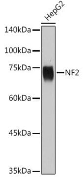
표적 설명
~70 kDa observed. 69.78/68.71 kDa (mouse/rat), 69.69/72.51/69.09/59.10/64.19/64.00/85.35 kDa (human isoform 1/2/3/4/5/6/8) calculated. Human spliced isoform 3, 4, 5, and 6 are also known as isoform II, delE2/3, delE3, and delE2, respectively. Uncharacterized bands may be observed in some lysate(s).
물리적 형태
Format: Purified
Protein G purified
Purified mouse IgG1 in buffer containing 0.1 M Tris-Glycine (pH 7.4), 150 mM NaCl with 0.05% sodium azide.
저장 및 안정성
Stable for 1 year at 2-8°C from date of receipt.
기타 정보
Concentration: Please refer to lot specific datasheet.
면책조항
Unless otherwise stated in our catalog or other company documentation accompanying the product(s), our products are intended for research use only and are not to be used for any other purpose, which includes but is not limited to, unauthorized commercial uses, in vitro diagnostic uses, ex vivo or in vivo therapeutic uses or any type of consumption or application to humans or animals.
적합한 제품을 찾을 수 없으신가요?
당사의 제품 선택기 도구.을(를) 시도해 보세요.
Storage Class Code
12 - Non Combustible Liquids
WGK
WGK 1
Flash Point (°F)
Not applicable
Flash Point (°C)
Not applicable
시험 성적서(COA)
제품의 로트/배치 번호를 입력하여 시험 성적서(COA)을 검색하십시오. 로트 및 배치 번호는 제품 라벨에 있는 ‘로트’ 또는 ‘배치’라는 용어 뒤에서 찾을 수 있습니다.
자사의 과학자팀은 생명 과학, 재료 과학, 화학 합성, 크로마토그래피, 분석 및 기타 많은 영역을 포함한 모든 과학 분야에 경험이 있습니다..
고객지원팀으로 연락바랍니다.