
추천 제품


생물학적 소스
rat
Quality Level
결합
unconjugated
항체 형태
purified antibody
항체 생산 유형
primary antibodies
클론
ER-TR5, monoclonal
종 반응성
human, mouse
포장
antibody small pack of 100 μg
기술
immunofluorescence: suitable
immunohistochemistry (formalin-fixed, paraffin-embedded sections): suitable
동형
IgM
에피토프 서열
Unknown
UniProt 수납 번호
배송 상태
2-8°C
타겟 번역 후 변형
unmodified
일반 설명
The thymus is considered as the primary lymphoid organ that is responsible for the generation and maturation of T cells. Thymic epithelial cells account for the majority of thymic stromal components. These cells can be divided into cortical and medullary thymic epithelial cells based on their localization within the thymus. Cortical thymic epithelial cells are essential for the positive selection of T cells, whereas the medullary thymic epithelial cells (mTEC) play a role in inducing negative selection of highly reactive T cells that are required for establishing central self-tolerance. In murine species, mTEC are further divided into mTEC low and mTEC high according to the expression levels of several maturation molecules, such as MHCII and CD80. mTEC low cells can serve as precursors for mature mTEC high cells. mTECs have also been classified into four major subsets, termed mTEC I-IV depending on their distinct transcriptional and molecular characteristics. mTEC I and II subpopulation appear early and can be detected at E18.5 and are most proliferative cells. mTEC III are detected in thymus at 4-weeks. mTEC IV are visible on day 6 in neonatal mice. The function of mTECs is dependent on their characteristic features such as ectopic expression of peripheral tissue restricted antigens and their master regulator-autoimmune regulator (Aire), expression of various chemokines and cytokines. Lymphotoxin β receptor (LTβR) is reported to be an essential regulator of multiple mTEC subsets within the mTEC low compartment. It controls thymic tolerance by regulating the frequency and makeup of intrathymic dendritic cells that are required for effective thymocyte negative selection. Clone ER-TR5 is shown to exclusively react with mTECs. (Ref.: Wang, HX., et al. (2020). Front. Immunol. 10; 3099; Lucas, B., et al. (2020). Nat. Commun. 11; Article 2198; Cosway, EJ., et al. (2017). J. Exp. Med. 214(11); 3183-3195; Van Vliet, E., et al. (1984). Eur. J. Immunol. 14(6); 524-529).
특이성
Clone ER-TR5 is a rat monoclonal antibody that detects Medullary epithelial cells.
면역원
Thymic stromal cells isolated from C3H mice.
애플리케이션
Quality Control Testing
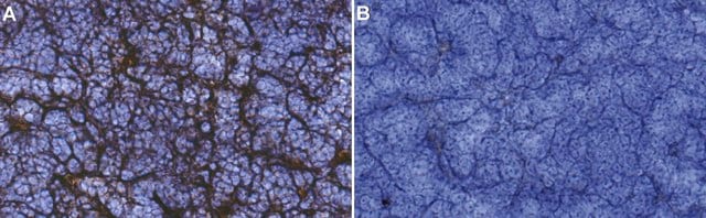
Evaluated by Immunohistochemistry in Mouse thymus tissue.
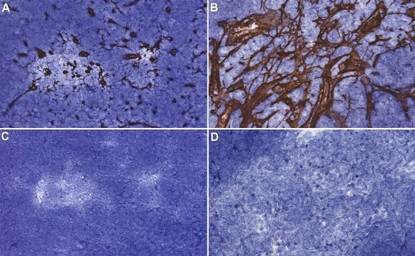
Immunohistochemistry Applications: 1:150 dilution of this antibody detected Medullary epithelium in Mouse thymus tissue sections.
Tested applications
Immunofluorescence Analysis: A representative lot detected Medullary epithelium in Immunofluorescence applications (Akiyama, T., et al. (2005). Science. 308(5719):248-51; Lei, Y., et al. (2011). J Exp Med. 208(2):383-94; Cosway, E.J., et al. (2017). J Exp Med. 214(11):3183-3195).
Immunohistochemistry Applications: A representative lot detected Medullary epithelium in Immunohistochemistry applications (Van Vliet, E., et al. (1984). Eur J Immunol. 14(6):524-9; Akiyama, T., et al. (2005). Science.;308(5719):248-51; Dooley, J., et al. (2006). J Immunol. 176(11):6484-90; Lei, Y., et al. (2011). J Exp Med. 208(2):383-94; Cosway, E.J., et al. (2017). J Exp Med. 214(11):3183-3195).
Note: Actual optimal working dilutions must be determined by end user as specimens, and experimental conditions may vary with the end user
Evaluated by Immunohistochemistry in Mouse thymus tissue.
Immunohistochemistry Applications: 1:150 dilution of this antibody detected Medullary epithelium in Mouse thymus tissue sections.
Tested applications
Immunofluorescence Analysis: A representative lot detected Medullary epithelium in Immunofluorescence applications (Akiyama, T., et al. (2005). Science. 308(5719):248-51; Lei, Y., et al. (2011). J Exp Med. 208(2):383-94; Cosway, E.J., et al. (2017). J Exp Med. 214(11):3183-3195).
Immunohistochemistry Applications: A representative lot detected Medullary epithelium in Immunohistochemistry applications (Van Vliet, E., et al. (1984). Eur J Immunol. 14(6):524-9; Akiyama, T., et al. (2005). Science.;308(5719):248-51; Dooley, J., et al. (2006). J Immunol. 176(11):6484-90; Lei, Y., et al. (2011). J Exp Med. 208(2):383-94; Cosway, E.J., et al. (2017). J Exp Med. 214(11):3183-3195).
Note: Actual optimal working dilutions must be determined by end user as specimens, and experimental conditions may vary with the end user
Anti-Medullary epithelium, clone ER-TR5, Cat. No. MABF2818, is a rat monoclonal antibody that detects Medullary epithelium and is tested for use in Immunohistochemistry and Immunofluorescence.
물리적 형태
Purified rat monoclonal antibody IgM in buffer containing 0.1 M Tris-Glycine (pH 7.4), 150 mM NaCl with 0.05% sodium azide.
저장 및 안정성
Recommended storage: +2°C to +8°C.
기타 정보
Concentration: Please refer to the Certificate of Analysis for the lot-specific concentration.
면책조항
Unless otherwise stated in our catalog or other company documentation accompanying the product(s), our products are intended for research use only and are not to be used for any other purpose, which includes but is not limited to, unauthorized commercial uses, in vitro diagnostic uses, ex vivo or in vivo therapeutic uses or any type of consumption or application to humans or animals.
적합한 제품을 찾을 수 없으신가요?
당사의 제품 선택기 도구.을(를) 시도해 보세요.
Storage Class Code
12 - Non Combustible Liquids
WGK
WGK 1
Flash Point (°F)
Not applicable
Flash Point (°C)
Not applicable
시험 성적서(COA)
제품의 로트/배치 번호를 입력하여 시험 성적서(COA)을 검색하십시오. 로트 및 배치 번호는 제품 라벨에 있는 ‘로트’ 또는 ‘배치’라는 용어 뒤에서 찾을 수 있습니다.
자사의 과학자팀은 생명 과학, 재료 과학, 화학 합성, 크로마토그래피, 분석 및 기타 많은 영역을 포함한 모든 과학 분야에 경험이 있습니다..
고객지원팀으로 연락바랍니다.