
추천 제품


생물학적 소스
mouse
Quality Level
항체 형태
purified antibody
항체 생산 유형
primary antibodies
클론
67A4, monoclonal
종 반응성
human
농도
0.5 mg/mL
기술
flow cytometry: suitable
immunofluorescence: suitable
inhibition assay: suitable
western blot: suitable
동형
IgG1κ
NCBI 수납 번호
UniProt 수납 번호
배송 상태
wet ice
타겟 번역 후 변형
unmodified
유전자 정보
human ... CDH1(999)
관련 카테고리
일반 설명
Cadherins are a family of transmembrane glycoproteins involved in Ca2+-dependent cell to cell interactions. The members of the family are differentially expressed in various tissues and function in the maintenance of tissue integrity, morphogenesis, and migration. Cadherins are divided into type I and type II subgroups. Type I cadherins include epithelial cadherin (E-cadherin), neural cadherin (N-cadherin), placental cadherin (P-cadherin) and retinal cadherin (R-cadherin). Type II include kidney cadherin (K-cadherin) and osteoblast cadherin (OB-cadherin). One of the best characterized cadherins is E-cadherin (epithelial cadherin or cadherin-1). It consists of calcium binding extracellular domains responsible for interactions with other E-cadherin molecules on neighboring cells and interaction with integrin alphaEbeta7. The cytoplasmic domain of E-cadherin is linked to the actin cytoskeleton through cytoplasmic catenin proteins, thus establishing a complex localized to adherens junctions. In carcinomas E-cadherin is frequently downregulated, which is consistent with its function of an invasion suppressor in normal epithelia.
면역원
Whole cells corresponding to human E-Cadherin.
애플리케이션
Flow Cytometry Analysis: 2 µg from a representative lot detected E-Cadherin in 1X10E6 A431 cells.
Flow Cytometry Analysis: A representative lot from an independent laboratory detected E-Cadherin in bone marrow cells (Burhing, H. J., et al. (1996). Leukemia. 10(1):106-116.)
Inhibition Analysis: A representative lot from an independent laboratory inhibited E-Cadherin-mediated adhesion of Langerhans to keratinocytes (Caberg, J. H., et al. (2008). Carcinogenesis. 29(7):1441-1447.).
Inhibition Analysis: A representative lot from an independent laboratory inhibits human embryoid body agglomeration (Dang, S. M., et al. (2004). Stem Cells. 22: 275-282.).
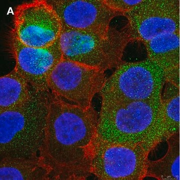
Immunofluorescence Analysis: A representative lot from an independent laboratory detected E-Cadherin in human thymus tissue (Kutlesa, S., et al. (2002). J Cell Sci. 115(Pt. 23):4505-4515.).
Western Blotting Analysis: A representative lot from an independent laboratory detected E-Cadherin in human thymus tissue lysate (Kutlesa, S., et al. (2002). J Cell Sci. 115(Pt. 23):4505-4515.).
Flow Cytometry Analysis: A representative lot from an independent laboratory detected E-Cadherin in bone marrow cells (Burhing, H. J., et al. (1996). Leukemia. 10(1):106-116.)
Inhibition Analysis: A representative lot from an independent laboratory inhibited E-Cadherin-mediated adhesion of Langerhans to keratinocytes (Caberg, J. H., et al. (2008). Carcinogenesis. 29(7):1441-1447.).
Inhibition Analysis: A representative lot from an independent laboratory inhibits human embryoid body agglomeration (Dang, S. M., et al. (2004). Stem Cells. 22: 275-282.).
Immunofluorescence Analysis: A representative lot from an independent laboratory detected E-Cadherin in human thymus tissue (Kutlesa, S., et al. (2002). J Cell Sci. 115(Pt. 23):4505-4515.).
Western Blotting Analysis: A representative lot from an independent laboratory detected E-Cadherin in human thymus tissue lysate (Kutlesa, S., et al. (2002). J Cell Sci. 115(Pt. 23):4505-4515.).
Research Category
Cell Structure
Cell Structure
Research Sub Category
ECM Proteins
ECM Proteins
This Anti-E-Cadherin Antibody, clone 67A4, Azide & Ascites Free is validated for use in western blotting, flow cytometry, inhibition & immunofluorescence for the detection of E-Cadherin.
품질
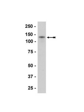
Evaluated by Western Blotting in A431 cell lysate.
Western Blotting Analysis: 0.25 µg/mL of this antibody detected E-Cadherin in 10 µg of A431 cell lysate.
Western Blotting Analysis: 0.25 µg/mL of this antibody detected E-Cadherin in 10 µg of A431 cell lysate.
표적 설명
~97 kDa observed
결합
Replaces: 04-1103
물리적 형태
Format: Purified
Protein G Purified
Purified mouse monoclonal IgG1κ in buffer containing PBS without preservatives.
저장 및 안정성
Stable for 1 year at -20°C from date of receipt.
Handling Recommendations: Upon receipt and prior to removing the cap, centrifuge the vial and gently mix the solution. Aliquot into microcentrifuge tubes and store at -20°C. Avoid repeated freeze/thaw cycles, which may damage IgG and affect product performance.
Handling Recommendations: Upon receipt and prior to removing the cap, centrifuge the vial and gently mix the solution. Aliquot into microcentrifuge tubes and store at -20°C. Avoid repeated freeze/thaw cycles, which may damage IgG and affect product performance.
면책조항
Unless otherwise stated in our catalog or other company documentation accompanying the product(s), our products are intended for research use only and are not to be used for any other purpose, which includes but is not limited to, unauthorized commercial uses, in vitro diagnostic uses, ex vivo or in vivo therapeutic uses or any type of consumption or application to humans or animals.
적합한 제품을 찾을 수 없으신가요?
당사의 제품 선택기 도구.을(를) 시도해 보세요.
Storage Class Code
12 - Non Combustible Liquids
WGK
WGK 2
Flash Point (°F)
Not applicable
Flash Point (°C)
Not applicable
시험 성적서(COA)
제품의 로트/배치 번호를 입력하여 시험 성적서(COA)을 검색하십시오. 로트 및 배치 번호는 제품 라벨에 있는 ‘로트’ 또는 ‘배치’라는 용어 뒤에서 찾을 수 있습니다.
자사의 과학자팀은 생명 과학, 재료 과학, 화학 합성, 크로마토그래피, 분석 및 기타 많은 영역을 포함한 모든 과학 분야에 경험이 있습니다..
고객지원팀으로 연락바랍니다.