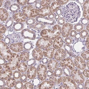
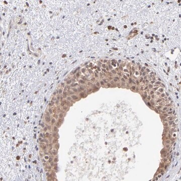
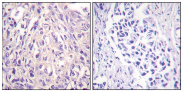
ABF117
Anti-IFIT1/p56 Antibody
serum, from rabbit
동의어(들):
Interferon-induced protein with tetratricopeptide repeats 1, IFIT-1, Glucocorticoid-attenuated response gene 16 protein, GARG-16, Interferon-induced 56 kDa protein, IFI-56K, P56
로그인조직 및 계약 가격 보기
모든 사진(2)
About This Item
UNSPSC 코드:
12352203
eCl@ss:
32160702
NACRES:
NA.41
추천 제품
생물학적 소스
rabbit
Quality Level
항체 형태
serum
항체 생산 유형
primary antibodies
클론
polyclonal
종 반응성
human, mouse
기술
flow cytometry: suitable
immunocytochemistry: suitable
western blot: suitable
NCBI 수납 번호
UniProt 수납 번호
배송 상태
wet ice
타겟 번역 후 변형
unmodified
유전자 정보
human ... IFIT1(3434)
일반 설명
IFIT1, also known as GARG-16, Glucocorticoid-attenuated response gene 16 protein or Interferon-induced 56 kDa protein, IFI-56K, or P56, and encoded by the gene lfit1/Garg16, Ifi56, Isg56, is an interferon-induced antiviral RNA-binding protein that specifically binds single-stranded RNA bearing a 5′-triphosphate group (PPP-RNA), thereby acting as a sensor of viral single-stranded RNAs and inhibiting expression of viral messenger RNAs. Single-stranded PPP-RNAs, which lack 2′-O-methylation of the 5′ cap and bear a 5′-triphosphate group instead, are specific from viruses, providing a molecular signature to distinguish between self and non-self mRNAs by the host during viral infection. IFIT1 directly binds PPP-RNA in a non-sequence-specific manner. Viruses evolved several ways to evade this restriction system such as encoding their own 2′-O-methylase for their mRNAs or by stealing host cap containing the 2′-O-methylation (cap snatching mechanism). IFIT1 is a component of an interferon-dependent multiprotein complex that is at least composed of IFIT1, IFIT2 and IFIT3 and interacts with EIF3F, RPL15, TMEM173 and EEF1A1 and is important in antiviral defense, and innate immune response. IFIT1 is localized to the cytoplasm and expressed by most cells, particularly in the immune system and epithelial cells.
면역원
GST-tagged recombinant protein corresponding to mouse IFIT1/p56.
애플리케이션
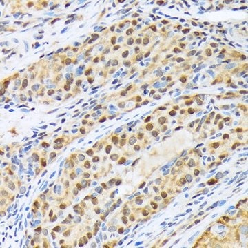
Detect IFIT1 using this rabbit polyclonal antibody, Anti-IFIT1/p56 Antibody validated for use in western blotting, ICC & Flow Cytometry.
Research Category
Inflammation & Immunology
Inflammation & Immunology
Research Sub Category
Inflammation & Autoimmune Mechanisms
Inflammation & Autoimmune Mechanisms
Western Blotting Analysis: A 1:1,000 dilution from a representative lot detected IFIT1/p56 in 10 µg of TF-1 and MOLT-4 cell lysates.
Western Blotting Analysis: A representative lot from an independent laboratory detected IFIT-1/p56 in HT1080 cell lysates transfected with mouse p56 vector and not in HT1080 cell lysates transfected with empty vector (Terenzi, F., et al. (2007). J Virol. 81(16):8656-8665.).
Western Blotting Analysis: A representative lot from an independent laboratory detected IFIT-1/p56 in IFN-Beta treated wild type Stat1 MEF cell lysates and not in IFN-beta treated Stat1 knockdown MEF cell lsyates (Fensterl, V., et al. (2008). J Virol. 82(22):11045-11053.).
Western Blotting Analysis: A representative lot from an independent laboratory detected IFIT-1/p56 in IFN-Beta treated wild type p56 MEF cell lysates and not in IFN-Beta treated p56 knockdown MEF cell lysates (Fensterl, V., et al. (2012). PLoS Pathog. 8(5):e1002712.).
Immunocytochemistry Analysis: A representative lot from an independent laboratory detected IFIT-1/p56 in IFN-Beta treated MEF cell lysates (Terenzi, F., et al. (2007). J Virol. 81(16):8656-8665.).
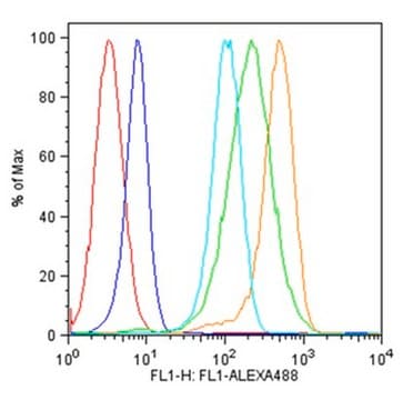
Flow Cytometry Analysis: A representative lot from an independent laboratory detected IFIT-1/p56 in bone marrow cells and splenocyotes upon injection with dsRNA, IFN-alpha, and VSV (Terenzi, F., et al. (2007). J Virol. 81(16):8656-8665.).
Flow Cytometry Analysis: A representative lot from an independent laboratory detected IFIT-1/p56 in IFN-Beta treated CD3+ mature T cells, and in IFN-Beta treated myeloid Dendritic cells, but not in IFN-Beta treated plasmacytoid Dendritic cells (Fensterl, V., et al. (2008). J Virol. 82(22):11045-11053.).
품질
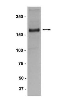
Evaluated by Western Blotting in mouse ovary tissue lysate.
Western Blotting Analysis: A 1:1,000 dilution of this antibody detected IFIT1/p56 in 10 µg of mouse ovary tissue lysate.
Western Blotting Analysis: A 1:1,000 dilution of this antibody detected IFIT1/p56 in 10 µg of mouse ovary tissue lysate.
표적 설명
~56 kDa observed. Uncharacterized band(s) may be observed in some cell lysates.
물리적 형태
Rabbit polyclonal serum containing 0.05% sodium azide.
Unpurified
저장 및 안정성
Stable for 1 year at -20°C from date of receipt.
Handling Recommendations: Upon receipt and prior to removing the cap, centrifuge the vial and gently mix the solution. Aliquot into microcentrifuge tubes and store at -20°C. Avoid repeated freeze/thaw cycles, which may damage IgG and affect product performance.
Handling Recommendations: Upon receipt and prior to removing the cap, centrifuge the vial and gently mix the solution. Aliquot into microcentrifuge tubes and store at -20°C. Avoid repeated freeze/thaw cycles, which may damage IgG and affect product performance.
면책조항
Unless otherwise stated in our catalog or other company documentation accompanying the product(s), our products are intended for research use only and are not to be used for any other purpose, which includes but is not limited to, unauthorized commercial uses, in vitro diagnostic uses, ex vivo or in vivo therapeutic uses or any type of consumption or application to humans or animals.
Not finding the right product?
Try our 제품 선택기 도구.
Storage Class Code
10 - Combustible liquids
WGK
WGK 1
시험 성적서(COA)
제품의 로트/배치 번호를 입력하여 시험 성적서(COA)을 검색하십시오. 로트 및 배치 번호는 제품 라벨에 있는 ‘로트’ 또는 ‘배치’라는 용어 뒤에서 찾을 수 있습니다.
Jayati Basu et al.
Nature immunology, 24(8), 1295-1307 (2023-07-21)
The transcription factor ThPOK (encoded by Zbtb7b) is well known for its role as a master regulator of CD4 lineage commitment in the thymus. Here, we report an unexpected and critical role of ThPOK as a multifaceted regulator of myeloid
자사의 과학자팀은 생명 과학, 재료 과학, 화학 합성, 크로마토그래피, 분석 및 기타 많은 영역을 포함한 모든 과학 분야에 경험이 있습니다..
고객지원팀으로 연락바랍니다.