추천 제품
생물학적 소스
rabbit
Quality Level
항체 형태
purified antibody
항체 생산 유형
primary antibodies
클론
polyclonal
종 반응성
human
기술
immunocytochemistry: suitable
western blot: suitable
동형
IgG
NCBI 수납 번호
UniProt 수납 번호
배송 상태
ambient
타겟 번역 후 변형
unmodified
유전자 정보
human ... CEP164(22897)
일반 설명
Centrosomal protein of 164 kDa (UniProt: Q9UPV0; also known as Cep164) is encoded by the CEP164 (also known as KIAA1052, NPHP15) gene (Gene ID: 22897) in human. Cep164 is expressed in several cell lines and it plays a role in microtubule organization and/or maintenance for the formation of primary cilia (PC), a microtubule-based structure that protrudes from the surface of epithelial cells. Cep164 localizes specifically to very distally located appendage structures on the mature centriole from which PC formation originates and it persists at centrioles throughout mitosis. Hence, it can serve as a marker for distal appendages on mature centrioles or basal bodies. Cep164 also plays a vital role in G2/M checkpoint and nuclear divisions. It is a key participant in the DNA damage-activated ATR/ATM signaling cascade and is required for the proper phosphorylation of H2AX, replication protein A, and check point kinases 1 and 2. Its phosphorylation at Ser186 is induced upon DNA-damage caused by treatment with IR irradiation, UV irradiation, hydroxyurea or amphidicolin. Mutations in CEP164 gene are known to cause nephronophthisis, a disease characterized by blindness in childhood, facial dysmorphism, bronchieclasis, liver failure and progression to end-stage renal failure.
특이성
This polyclonal antibody detects Centrosomal protein of 164 kDa in human. It targets an epitope within the first 298 amino acids from the N-terminal region.
면역원
Epitope: N-terminus
His-taggged recombinant fragment of the first 298 amino acids from human Centrosomal protein of 164 kDa (CEP164).
애플리케이션
Anti-CEP164, Cat. No. ABE2621, is a highly specific rabbit polyclonal antibody that targets Centrosomal protein of 164 kDa and has been tested in Immunocytochemistry and Western Blotting.
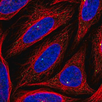
Immunocytochemistry Analysis: A representative lot detected CEP164 in U2OS cells and hTERT-RPE1 cells that were serum starved for 48 hours (Graser, S., et. al. (2007). J Cell Biol. 179(2):321-30; Sonnen, K.F., et. al. (2012). Biol Open. 1(10):965-76).
Western Blotting Analysis: A representative lot detected CEP164 in total lysates of HeLa S3, U2OS, and 293T cells as well as 293T cells overexpressing myc-Cep164 (Graser, S., et. al. (2007). J Cell Biol. 179(2):321-30).
Western Blotting Analysis: A representative lot detected CEP164 in total lysates of HeLa S3, U2OS, and 293T cells as well as 293T cells overexpressing myc-Cep164 (Graser, S., et. al. (2007). J Cell Biol. 179(2):321-30).
Research Category
Epigenetics & Nuclear Function
Epigenetics & Nuclear Function
품질
Evaluated by Western Blotting in U2OS cell lysate.
Western Blotting Analysis: A 1:500 dilution of this antibody detected CEP164 in 10 µg of U2OS cell lysate.
Western Blotting Analysis: A 1:500 dilution of this antibody detected CEP164 in 10 µg of U2OS cell lysate.
표적 설명
~200 kDa observed; 164.31 kDa calculated. Uncharacterized bands may be observed in some lysate(s).
물리적 형태
Format: Purified
Protein A purified
Purified rabbit polyclonal antibody in buffer containing 0.1 M Tris-Glycine (pH 7.4), 150 mM NaCl with 0.05% sodium azide.
저장 및 안정성
Stable for 1 year at 2-8°C from date of receipt.
기타 정보
Concentration: Please refer to lot specific datasheet.
면책조항
Unless otherwise stated in our catalog or other company documentation accompanying the product(s), our products are intended for research use only and are not to be used for any other purpose, which includes but is not limited to, unauthorized commercial uses, in vitro diagnostic uses, ex vivo or in vivo therapeutic uses or any type of consumption or application to humans or animals.
적합한 제품을 찾을 수 없으신가요?
당사의 제품 선택기 도구.을(를) 시도해 보세요.
Storage Class Code
12 - Non Combustible Liquids
WGK
WGK 1
시험 성적서(COA)
제품의 로트/배치 번호를 입력하여 시험 성적서(COA)을 검색하십시오. 로트 및 배치 번호는 제품 라벨에 있는 ‘로트’ 또는 ‘배치’라는 용어 뒤에서 찾을 수 있습니다.
Phillip Scott et al.
The Journal of cell biology, 222(12) (2023-09-29)
Polo-like kinase 4 (PLK4) is a key regulator of centriole biogenesis, but how PLK4 selects a single site for procentriole assembly remains unclear. Using ultrastructure expansion microscopy, we show that PLK4 localizes to discrete sites along the wall of parent
Thao P Phan et al.
Genes & development, 36(11-12), 718-736 (2022-07-01)
Centrosomes are microtubule-organizing centers comprised of a pair of centrioles and the surrounding pericentriolar material. Abnormalities in centriole number are associated with cell division errors and can contribute to diseases such as cancer. Centriole duplication is limited to once per
Olivier Mercey et al.
Nature cell biology, 21(12), 1544-1552 (2019-12-04)
Multiciliated cells (MCCs) amplify large numbers of centrioles that convert into basal bodies, which are required for producing multiple motile cilia. Most centrioles amplified by MCCs grow on the surface of organelles called deuterosomes, whereas a smaller number grow through
자사의 과학자팀은 생명 과학, 재료 과학, 화학 합성, 크로마토그래피, 분석 및 기타 많은 영역을 포함한 모든 과학 분야에 경험이 있습니다..
고객지원팀으로 연락바랍니다.