콘텐츠로 건너뛰기
Merck
모든 사진(2)

문서

AB6000

Sigma-Aldrich

Anti-TIMP-3 Antibody, CT

from rabbit, purified by affinity chromatography

동의어(들):

MIG-5, TIMP metallopeptidase inhibitor 3, Tissue inhibitor of metalloproteinases 3

로그인조직 및 계약 가격 보기


About This Item

UNSPSC 코드:
12352203
eCl@ss:
32160702
NACRES:
NA.41

생물학적 소스

rabbit

Quality Level

항체 형태

affinity isolated antibody

항체 생산 유형

primary antibodies

클론

polyclonal

정제법

affinity chromatography

종 반응성

horse, human, rat

종 반응성(상동성에 의해 예측)

pig (based on 100% sequence homology), primate (based on 100% sequence homology), equine (based on 100% sequence homology), canine (based on 100% sequence homology), monkey (based on 100% sequence homology), mouse (95% homology), bovine (based on 100% sequence homology)

기술

immunocytochemistry: suitable
western blot: suitable

NCBI 수납 번호

UniProt 수납 번호

배송 상태

wet ice

타겟 번역 후 변형

unmodified

유전자 정보

human ... TIMP3(7078)

일반 설명

Tissue inhibitor of metalloproteinases 3 (TIMP-3) is a member of the TIMP family. The TIMPs are so named because they are slow, tight binding endogenous inhibitors of the Matrix Metalloproteinases (MMPs). The four TIMPs have different inhibition constants for the different MMPs studied. TIMPs have been shown to have activity against the ADAMs family of proteinases. TIMP-3 is an efficient "sheddase" inhibitor, inhibiting ADAM-17 (TACE) at the low nanomolar levels seen with MMP inhibition. The other ADAMs proteinases have not yet been assayed for TIMP inhibition, but TIMP-2 seems to be much less active on ADAM-17 than isTIMP-3. TIMP-3 is thought to be constitutively produced by many cell types and is inducible in others. The TIMP-3 localization differs from the other 3 TIMPs, and is thought to be primarily deposited into the extracellular matrix.

특이성

The antibody recognizes TIMP-3 at the C-terminus. Does not appear to cross react with TIMP-1 or TIMP-2.

면역원

Epitope: C-terminus
KLH-conjugated linear peptide corresponding to human TIMP-3 at the C-terminus.

애플리케이션

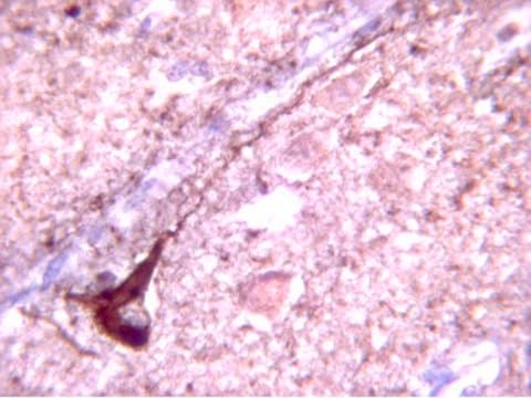
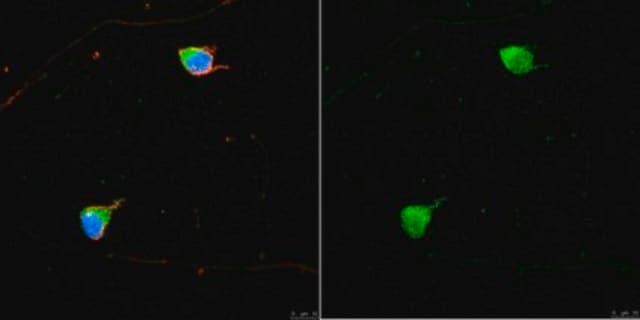
Immunocytochemistry Analysis: 1:500 dilution from a previous lot detected TIMP-3 in C6 cells.
Research Category
Cell Structure
Research Sub Category
MMPs & TIMPs
This Anti-TIMP-3 Antibody, C-terminus is validated for use in WB, IC for the detection of TIMP-3.

품질

Evaluated by Western Blot using an HL-60 cell lysate supplemented with PMA-conditioned media.

Western Blot Analysis: 0.5 µg/ml of this antibody detected TIMP-3 on 10 µg of HL-60 in PMA-conditioned media.

표적 설명

Bands may be observed for reduced protein bands at ~ 24 kDa (unglycosylated) and ~ 30 kDa (glycosylated). Additional TIMP-3 bands MAY be visible at 50 kDa (dimer), 12 kDa, and 15 kDa (breakdown products) and sample dependent.

결합

Replaces: AB802

물리적 형태

Affinity purified
Purified rabbit polyclonal in buffer containing 0.1 M Tris-Glycine (pH 7.4, 150 mM NaCl) with 0.05% sodium azide.

저장 및 안정성

Stable for 1 year at 2-8°C from date of receipt.

분석 메모

Control
HL-60 cell lysate in PMA-conditioned media.

기타 정보

Concentration: Please refer to the Certificate of Analysis for the lot-specific concentration.

면책조항

Unless otherwise stated in our catalog or other company documentation accompanying the product(s), our products are intended for research use only and are not to be used for any other purpose, which includes but is not limited to, unauthorized commercial uses, in vitro diagnostic uses, ex vivo or in vivo therapeutic uses or any type of consumption or application to humans or animals.
Not finding the right product?  

Try our 제품 선택기 도구.

Storage Class Code

12 - Non Combustible Liquids

WGK

WGK 1

Flash Point (°F)

Not applicable

Flash Point (°C)

Not applicable


시험 성적서(COA)

제품의 로트/배치 번호를 입력하여 시험 성적서(COA)을 검색하십시오. 로트 및 배치 번호는 제품 라벨에 있는 ‘로트’ 또는 ‘배치’라는 용어 뒤에서 찾을 수 있습니다.

이 제품을 이미 가지고 계십니까?

문서 라이브러리에서 최근에 구매한 제품에 대한 문서를 찾아보세요.

문서 라이브러리 방문

Ioannis Kanakis et al.
Arthritis & rheumatology (Hoboken, N.J.), 71(4), 571-582 (2018-11-01)
Cartilage destruction in osteoarthritis (OA) is mediated mainly by matrix metalloproteinases (MMPs) and ADAMTS. The therapeutic candidature of targeting aggrecanases has not yet been defined in joints in which spontaneous OA arises from genetic susceptibility, as in the case of
Basic-science observations explain how outer retinal hyperreflective foci predict drusen regression and geographic atrophy in age-related macular degeneration.
Qitao Zhang et al.
Eye (London, England), 36(5), 1115-1118 (2021-08-22)
Jung Hyun Park et al.
Journal of personalized medicine, 12(5) (2022-05-29)
Tissue inhibitor of metalloproteinase-3 (TIMP-3) is a component of the extracellular environment and is suggested to play an indirect role in regulating Aβ production and the pathophysiology of Aβ deposition in brains. However, studies on the amount of TIMP-3 in
Qitao Zhang et al.
Experimental eye research, 178, 212-222 (2018-10-20)
The daily shedding and renewal of photoreceptor outer segments (OS) is critical for maintaining vision. This process relies on the efficient uptake, degradation, and sorting of shed OS material by the retinal pigment epithelium (RPE). Poor OS degradation has been
Qitao Zhang et al.
Investigative ophthalmology & visual science, 60(10), 3468-3479 (2019-08-14)
The accumulation of undigestible autofluorescent material (UAM), termed lipofuscin in vivo, is a hallmark of aged RPE. Lipofuscin derives, in part, from the incomplete degradation of phagocytized photoreceptor outer segments (OS). Whether this accumulated waste is toxic is unclear. We

자사의 과학자팀은 생명 과학, 재료 과학, 화학 합성, 크로마토그래피, 분석 및 기타 많은 영역을 포함한 모든 과학 분야에 경험이 있습니다..

고객지원팀으로 연락바랍니다.