05-338
Anti-Fas Antibody (human, neutralizing), clone ZB4
clone ZB4, Upstate®, from mouse
동의어(들):
APO-1 cell surface antigen, Apo-1 antigen, Apoptosis-mediating surface antigen FAS, CD95 antigen, FASLG receptor, Fas (TNF receptor superfamily, member 6), Fas AMA, Fas antigen, apoptosis antigen 1, tumor necrosis factor receptor superfamily member 6, tu
로그인조직 및 계약 가격 보기
모든 사진(1)
About This Item
UNSPSC 코드:
12352203
eCl@ss:
32160702
NACRES:
NA.41
추천 제품
관련 카테고리
일반 설명
Fas (also known as Apo-1 and CD95) is a 40-50 kDa cell surface glycoprotein and a member of the TNF receptor family. Fas is an apotosis signalling molecule that forms the death inducing signalling complex (DISC) upon binding by the Fas ligand trimer on an adjacent cell. The Fas receptor also trimerizes, after which it is internalized and can then bind to the adaptor molecule FADD. FADD facilitates binding of caspase-8. which is activated to induce caspase-8 mediated apoptosis. Fas also plays a role in T cell mediated cytotoxicity and T cell development. Fas is found in the thymus, liver, heart, and ovary, where it is expressed on a minority of resting T and B cells and monocytes. The antagonistic ZB4 clone anti-Fas monoclonal has been used to block the apoptosis-inducing activity of the Anti-Fas CH-11 clone in multiple publications.
This product does not induce apoptosis in cell culture.
특이성
This antibody does not recognize TNF and mouse Fas.
This antibody recognizes the human cell surface antigen Fas expressed in various human cells, including myeloid cells, T lympho-blastoid cells and diploid fibroblasts. It does not induce apoptosis in cell culture.
면역원
Recombinant human Fas.
애플리케이션
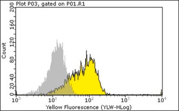
Anti-Fas Antibody (human, neutralizing), clone ZB4 detects level of Fas & has been published & validated for use in WB, FC, NEUT.
Flow Cytometry: A previous lot of this antibody used 5-20 μg/mL. Western Blot: A previous lot of this antibody used 5 μg/mL.
Research Category
Apoptosis & Cancer
Apoptosis & Cancer
Research Sub Category
Apoptosis - Additional
Apoptosis - Additional
품질
Evaluated by neutralization of apoptosis induced by anti-Fas (human, activating), clone CH11 (Catalog # 05-201) treatment of human Jurkat cells.
Neutralization: 10-500 ng/mL of this antibody was incubated for one hour with human Fas-transfected cells and inhibited the anti-Fas (human, activating), clone CH11 (Catalog # 05-201). Cell viability was assessed using a WST-1 assay
Neutralization: 10-500 ng/mL of this antibody was incubated for one hour with human Fas-transfected cells and inhibited the anti-Fas (human, activating), clone CH11 (Catalog # 05-201). Cell viability was assessed using a WST-1 assay
표적 설명
45 kDa
물리적 형태
Ammonium sulfate and purified mouse monoclonal IgG1 in PBS containing 50% glycerol. Store at -20°C.
Format: Purified
Protein A purified
저장 및 안정성
Stable for 1 year at from date of receipt.
분석 메모
Control
Various human cells
Various human cells
기타 정보
Concentration: Please refer to the Certificate of Analysis for the lot-specific concentration.
법적 정보
UPSTATE is a registered trademark of Merck KGaA, Darmstadt, Germany
면책조항
Unless otherwise stated in our catalog or other company documentation accompanying the product(s), our products are intended for research use only and are not to be used for any other purpose, which includes but is not limited to, unauthorized commercial uses, in vitro diagnostic uses, ex vivo or in vivo therapeutic uses or any type of consumption or application to humans or animals.
Not finding the right product?
Try our 제품 선택기 도구.
Storage Class Code
10 - Combustible liquids
WGK
WGK 2
시험 성적서(COA)
제품의 로트/배치 번호를 입력하여 시험 성적서(COA)을 검색하십시오. 로트 및 배치 번호는 제품 라벨에 있는 ‘로트’ 또는 ‘배치’라는 용어 뒤에서 찾을 수 있습니다.
Stimulated human lamina propria T cells manifest enhanced Fas-mediated apoptosis
Boirivant, M., et al
The Journal of Clinical Investigation, 98, 2616-2622 (1996)
E Lacana et al.
Nature medicine, 5(5), 542-547 (1999-05-06)
Programmed cell death is a process required for the normal development of an organism. One of the best understood apoptotic pathways occurs in T lymphocytes and is mediated by Fas/Fas ligand (FasL) interaction. During studies of apoptosis induced by T
Networked T cell death following macrophage infection by Mycobacterium tuberculosis.
Macdonald, SH; Woodward, E; Coleman, MM; Dorris, ER; Nadarajan, P; Chew, WM; McLaughlin et al.
Testing null
Molecular determinants of apoptosis induced by the cytotoxic ribonuclease onconase: evidence for cytotoxic mechanisms different from inhibition of protein synthesis.
M S Iordanov, O P Ryabinina, J Wong, T H Dinh, D L Newton, S M Rybak, B E Magun
Cancer Research null
D Meley et al.
Cell death & disease, 1, e41-e41 (2011-03-03)
Medulloblastoma (MB) is an embryonic brain tumour that arises in the cerebellum. Using several MB cell lines, we have demonstrated that the chemotherapeutic drug etoposide induces a p53- and caspase-dependent cell death. We have observed an additional caspase-independent cell death
자사의 과학자팀은 생명 과학, 재료 과학, 화학 합성, 크로마토그래피, 분석 및 기타 많은 영역을 포함한 모든 과학 분야에 경험이 있습니다..
고객지원팀으로 연락바랍니다.


