Skip to Content
Merck
All Photos(1)

Key Documents

S2177

Sigma-Aldrich

Anti-Synaptotagmin antibody produced in rabbit

IgG fraction of antiserum, buffered aqueous solution

Sign Into View Organizational & Contract Pricing


About This Item

MDL number:
UNSPSC Code:
12352203
NACRES:
NA.46

biological source

rabbit

conjugate

unconjugated

antibody form

IgG fraction of antiserum

antibody product type

primary antibodies

clone

polyclonal

form

buffered aqueous solution

mol wt

antigen 65 kDa

species reactivity

rat

technique(s)

microarray: suitable
western blot: 1:5,000 using extract of rat brain membrane fraction

UniProt accession no.

shipped in

dry ice

storage temp.

−20°C

target post-translational modification

unmodified

Gene Information

human ... SYT1(6857)
mouse ... Syt1(20979)
rat ... Syt1(25716)

General description

Synaptotagmin (Syt, p65) is an abundant synaptic vesicle (SV) membrane protein. It is characterized by a short intravesicular N-terminal domain, a single transmembrane region and two copies of highly conserved internal repeats, known as the C2A and C2B domains, which are homologous to the C2 regulatory region of protein kinase C (PKC) in the cytoplasmic domain. At least eight different isoforms of synaptotagmin (SytI-VIII) are expressed in the brain, four of which (Syt IV, V, VII and VIII) are also expressed in non-neuronal tissues.

Specificity

The sequence is highly conserved among species (SytI) and is not found in other known synaptotagmin isoforms (SytII-VIII).

Immunogen

synthetic peptide corresponding to N-terminus of synaptotagmin I (SytI) of rat origin (amino acids 1-16 with C-terminally added lysine), conjugated to KLH.

Application

Anti-Synaptotagmin antibody produced in rabbit has been used:
  • for cell surface labelling of synaptotagmin I (SytI)
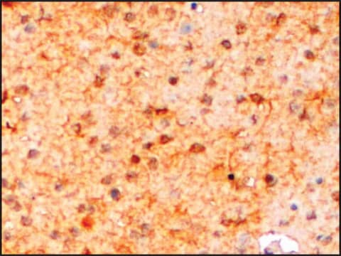
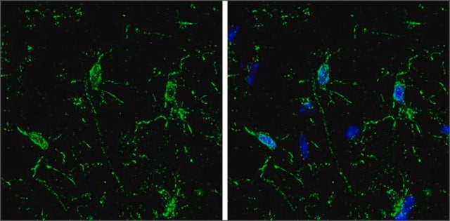
  • in immunofluorescence microscopy
  • in western blotting

Biochem/physiol Actions

Synaptotagmin binds Ca2+ phospholipids with high affinity and has a central role in Ca2+ regulated neurotransmitter release. Synaptotagmin functions as a Ca2+ sensor and is required for efficient exocytosis, particularly in the vesicle docking and/or fusion step with the plasma membrane. Ca2+ influx triggers synaptotagmin to interact with either syntaxin or SNAP-25 and the cytoplasmic domain of neurexin leading to fusion and exocytosis. Mutations or deletion of synaptotagmin result in severely impaired Ca2+ triggered neurotransmitter release. Synapses of SytI knockout mice lack the fast-component of Ca2+ dependent neurotransmitter release, but exhibit no changes in the slow, Ca2+ independent component of synaptic vesicle exocytosis.

Physical form

Solution in 0.01 M phosphate buffered saline, pH 7.4, containing 15 mM sodium azide.

Disclaimer

Unless otherwise stated in our catalog or other company documentation accompanying the product(s), our products are intended for research use only and are not to be used for any other purpose, which includes but is not limited to, unauthorized commercial uses, in vitro diagnostic uses, ex vivo or in vivo therapeutic uses or any type of consumption or application to humans or animals.

Not finding the right product?  

Try our Product Selector Tool.

Storage Class Code

12 - Non Combustible Liquids

WGK

nwg

Flash Point(F)

Not applicable

Flash Point(C)

Not applicable


Certificates of Analysis (COA)

Search for Certificates of Analysis (COA) by entering the products Lot/Batch Number. Lot and Batch Numbers can be found on a product’s label following the words ‘Lot’ or ‘Batch’.

Already Own This Product?

Find documentation for the products that you have recently purchased in the Document Library.

Visit the Document Library

The C2B domain of synaptotagmin is a Ca2+ sensing module essential for exocytosis
Desai RC, et al.
The Journal of Cell Biology, 150(5), 1125-1136 (2000)
Synaptotagmin in Ca2+dependent exocytosis: dynamic action in a flash
Tokuoka H and Goda Y
Neuron, 38(4), 521-524 (2003)
Alzheimer-related decrease in CYFIP2 links amyloid production to tau hyperphosphorylation and memory loss
Tiwari SS, et al.
Brain, 139(10), 2751-2751 (2016)
Scott W Messenger et al.
American journal of physiology. Gastrointestinal and liver physiology, 305(6), G439-G452 (2013-07-23)
Zymogen granule (ZG) formation in acinar cells involves zymogen cargo sorting from trans-Golgi into immature secretory granules (ISGs). ISG maturation progresses by removal of lysosomal membrane and select content proteins, which enter endosomal intermediates prior to their apical exocytosis. Constitutive
The C2 domains of synaptotagmin-partners in exocytosis
Bai J and Chapman ER
Trends in Biochemical Sciences, 29(3), 143-151 (2004)

Our team of scientists has experience in all areas of research including Life Science, Material Science, Chemical Synthesis, Chromatography, Analytical and many others.

Contact Technical Service