Skip to Content
Merck
All Photos(1)

Documents

252M-1

Sigma-Aldrich

Fascin (55k-2) Mouse Monoclonal Antibody

Sign Into View Organizational & Contract Pricing


About This Item

UNSPSC Code:
12352203
NACRES:
NA.41

biological source

mouse

Quality Level

100
500

conjugate

unconjugated

antibody form

culture supernatant

antibody product type

primary antibodies

clone

55k-2, monoclonal

description

For In Vitro Diagnostic Use in Select Regions (See Chart)

form

buffered aqueous solution

species reactivity

human

packaging

vial of 0.1 mL concentrate (252M-14)
vial of 0.5 mL concentrate (252M-15)
bottle of 1.0 mL predilute (252M-17)
vial of 1.0 mL concentrate (252M-16)
bottle of 7.0 mL predilute (252M-18)

manufacturer/tradename

Cell Marque

technique(s)

immunohistochemistry (formalin-fixed, paraffin-embedded sections): 1:100-1:500

isotype

IgG1

control

Hodgkin lymphoma

shipped in

wet ice

storage temp.

2-8°C

visualization

cytoplasmic

Gene Information

human ... FSCN1(6624)

General description

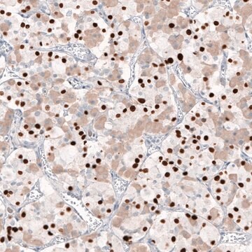
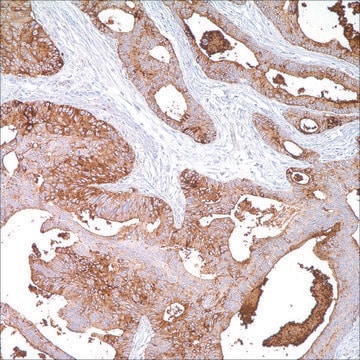
Anti-fascin is a very sensitive marker for Reed-Sternberg cells and variants in nodular sclerosis, mixed cellularity, and lymphocyte depletion Hodgkin disease. It is uniformly negative in lymphoid cells, plasma cells, and myeloid cells. Anti-fascin is positive in dendritic cells. This marker may be helpful to distinguish between Hodgkin lymphoma and non-Hodgkin lymphoma in difficult cases. Also, the lack of expression of fascin in the neoplastic follicles in follicular lymphoma can be helpful in distinguishing these lymphomas from reactive follicular hyperplasia in which the number of follicular dendritic cells is normal or increased.

Quality


IVD

IVD

IVD

RUO

Linkage

Fascin Positive Control Slides, Product No. 252S, are available for immunohistochemistry (formalin-fixed, paraffin-embedded sections).

Physical form

Solution in Tris Buffer, pH 7.3-7.7, with 1% BSA and <0.1% Sodium Azide

Preparation Note

Download the IFU specific to your product lot and formatNote: This requires a keycode which can be found on your packaging or product label.

Other Notes

For Technical Service please contact: 800-665-7284 or email: service@cellmarque.com

Legal Information

Cell Marque is a trademark of Merck KGaA, Darmstadt, Germany

Not finding the right product?  

Try our Product Selector Tool.


Certificates of Analysis (COA)

Search for Certificates of Analysis (COA) by entering the products Lot/Batch Number. Lot and Batch Numbers can be found on a product’s label following the words ‘Lot’ or ‘Batch’.

Already Own This Product?

Find documentation for the products that you have recently purchased in the Document Library.

Visit the Document Library

W Hu et al.
Clinical & experimental metastasis, 18(1), 83-88 (2001-02-24)
Fascin bundles actin microfilaments within dynamic cellular structures such as microspikes, stress fibers and membrane ruffles. Fascin overexpression induces membrane protrusions and increased cell motility, and is highly expressed in various transformed cells, and in specialized normal cells including neuronal
Giuseppe Pelosi et al.
Lung cancer (Amsterdam, Netherlands), 42(2), 203-213 (2003-10-22)
Immunoreactivity for fascin, an actin-bundling protein related to cell motility, has been reported in breast, ovary, pancreas, skin, and non-small cell carcinomas, and associated with more advanced disease stage and poorer prognosis. Data on pulmonary neuroendocrine (NE) tumors, however, are
Werner Kempf et al.
Journal of cutaneous pathology, 29(5), 295-300 (2002-07-09)
CD30-positive cutaneous lymphoproliferative disorders (LPD) represent a spectrum of diseases ranging from low-grade (lymphomatoid papulosis; LyP) to high-grade (pleomorphic and anaplastic large-cell lymphoma; PTL, ALCL) with overlapping morphologic and immunophenotypic features. The common phenotypic hallmark is the expression of CD30-antigen
G S Pinkus et al.
The American journal of pathology, 150(2), 543-562 (1997-02-01)
Immunohistochemical localization of human fascin, a distinct 55-kd actin-bundling protein, was determined for a wide variety of lymphoid tissues (364 specimens total). In non-neoplastic tissues, reactivity was highly selective and localized predominantly in dendritic cells. In the thymus, this protein
M D Kraus et al.
The American Journal of dermatopathology, 23(2), 104-111 (2001-04-04)
The non-Langerhans histiocytoses, a nosologic category to which juvenile xanthogranuoma (JXG) belongs, represent a heterogenous collection of disorders related to the monocyte/macrophage lineage. The dermal dendrocyte was previously proposed as the cell of origin for JXG on the basis of

Our team of scientists has experience in all areas of research including Life Science, Material Science, Chemical Synthesis, Chromatography, Analytical and many others.

Contact Technical Service