Monoclonal Anti-TRF1 (mouse IgG1 isotype) is derived from the TRF-78 hybridoma produced by the fusion of mouse myeloma cells and splenocytes from mice immunized with a human TRF1 protein produced in baculovirus. TRF1 and TRF2 (TTAGGG repeat binding factors) are two major proteins that bind to human telomers. TRF1 has a DNA binding domain with high homology to the Myb family of transcription factors. Unlike the Myb family that contains only one DNA binding motif, TRF1 has multiple of this motif.
Specificity
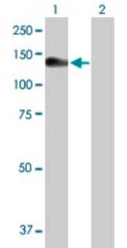
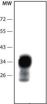
Monoclonal Anti-TRF1 recognizes human TRF1.
Immunogen
human TRF1 protein produced in baculovirus.
Application
Monoclonal Anti-TRF1 antibody produced in mouse has been used in Western blotting and enzyme linked immunosorbent assay (ELISA).
Biochem/physiol Actions
TRF1 (TTAGGG repeat binding factor 1) has a negative effect on the length of the telomer. Overexpression of TRF1 in cancer cells that contain telomerase activity, causes the shortening of the length of their telomers. While inhibition of TRF1 causes the elongation of telomers. It was shown that the level of TRF1 in the cells does not affect the expression of the telomerase protein. This suggests that TRF1 may act directly on the activity of the telomerase protein. Tankyrase is a protein that interacts with TRF1 and its C-terminal region is homologous to poly (adenosine dinucleotide phosphate (ADP)-ribose) polymerase (PARP). In response to DNA damage, the PARP protein mediates ADP-ribose polymers of protein acceptors. In vitro studies have shown that tankyrase is responsible for that polyribosylation of TRF1, which in turn abolishes its ability to bind telomers.
Physical form
Solution in 0.01 M phosphate buffered saline, pH 7.4, and 15 mM sodium azide.
Disclaimer
Unless otherwise stated in our catalog or other company documentation accompanying the product(s), our products are intended for research use only and are not to be used for any other purpose, which includes but is not limited to, unauthorized commercial uses, in vitro diagnostic uses, ex vivo or in vivo therapeutic uses or any type of consumption or application to humans or animals.
Mitochondria change their shape through fusion and fission in order to adapt to their metabolic milieu. Mitofusin-2 (MFN2) is a key regulatory protein in this process, mediating mitochondrial fusion and interaction with endoplasmic reticulum. Targeted deletion of Mfn2 in oocytes
Bortezomib-mediated down-regulation of telomerase and disruption of telomere homeostasis contributes to apoptosis of malignant cells
Bortezomib inhibits the ubiquitin/proteasome pathway to achieve its anti-cancer effect and its well characterized activity is the NF-κB inhibition through which the anti-apoptotic bcl-2 expression is down-regulated and apoptosis is subsequently induced. However, the downstream molecular targets of bortezomib are
Our team of scientists has experience in all areas of research including Life Science, Material Science, Chemical Synthesis, Chromatography, Analytical and many others.