Skip to Content
Merck
All Photos(3)

Key Documents

N3038

Sigma-Aldrich

Anti-Nanog antibody, Mouse monoclonal

clone NNG-811, purified from hybridoma cell culture

Synonym(s):

Anti-NANOG/STM1

Sign Into View Organizational & Contract Pricing


About This Item

UNSPSC Code:
12352203
NACRES:
NA.41

biological source

mouse

conjugate

unconjugated

antibody form

purified immunoglobulin

antibody product type

primary antibodies

clone

NNG-811, monoclonal

form

buffered aqueous solution

mol wt

~40 kDa

species reactivity

human

packaging

antibody small pack of 25 μL

concentration

~2 mg/mL

technique(s)

immunocytochemistry: suitable
immunoprecipitation (IP): suitable
indirect ELISA: suitable
western blot: 4-8 μg/mL

isotype

IgG1

UniProt accession no.

shipped in

dry ice

storage temp.

−20°C

target post-translational modification

unmodified

Gene Information

human ... NANOG(79923)

General description

Anti-Nanog antibody, Mouse monoclonal (mouse IgG1 isotype) is derived from the hybridoma NNG-811 produced by the fusion of mouse myeloma cells and splenocytes from BALB/c mice immunized with recombinant human Nanog. The human Nanog gene product is a 305 amino acid, 35 kDa protein with a tryptophan repeat domain and two C-terminal trans-activating domains. It is primarily located in the nucleus.
Nanog is a homeodomain transcription factor expressed in cells of the inner cell mass of early embryos, in embryonic stem cells (ESC), germ cells and in stem/progenitor cells of some tissues. Together with Oct-4, Sox-2, and Rex-1, it is a molecular marker for pluripotent cells and for undifferentiated stem cells. This protein is crucial for the maintenance of pluripotency of ESCs and is down-regulated when ESCs differentiate.
Nanog controls the expression of many ESC genes together with other stem cell transcription factors like Oct-4 and Sox-2. Nanog targets both repressor and activator complexes to regulatory regions of hundreds of genes in the genome. Expression of nanog can be detected primarily in germ cell tumors and in tumors of other cell types. Nanog is an important marker for Seminomas, testicular carcinomas, teratocarcinomas, and germ cell-like tumors in various tissues. Furthermore, it was shown to transform NIH3T3 cells.

Specificity

Monoclonal Anti-Nanog reacts specifically with human Nanog.

Immunogen

recombinant human Nanog.

Application

Anti-Nanog antibody, Mouse monoclonal has been used in:
  • enzyme-linked immunosorbent assay (ELISA)
  • immunoblotting
  • immunocytochemistry
  • flow cytometric analysis
  • immunoprecipitation
  • immunofluorescence

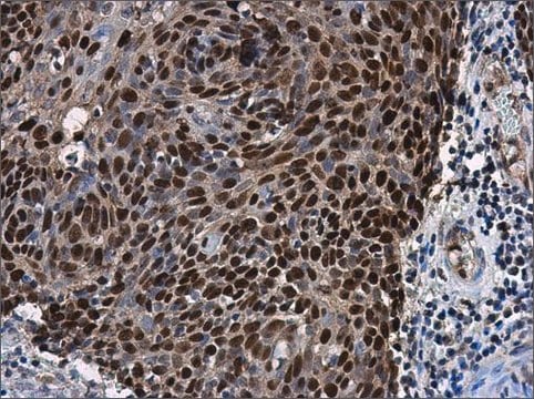
Monoclonal Anti-Nanog antibody is suitable for use in ELISA, immunoblotting (~ 40 kDa), immunocytochemistry and immunoprecipitation.
Immunoblotting: a working antibody concentration of 2-4 mg/mL is recommended using extracts of NT2 cells.

Physical form

Solution in 0.01 M phosphate buffered saline, pH 7.4, containing 15 mM sodium azide.

Disclaimer

Unless otherwise stated in our catalog or other company documentation accompanying the product(s), our products are intended for research use only and are not to be used for any other purpose, which includes but is not limited to, unauthorized commercial uses, in vitro diagnostic uses, ex vivo or in vivo therapeutic uses or any type of consumption or application to humans or animals.

Not finding the right product?  

Try our Product Selector Tool.

Storage Class Code

10 - Combustible liquids

WGK

WGK 3

Flash Point(F)

Not applicable

Flash Point(C)

Not applicable

Personal Protective Equipment

dust mask type N95 (US), Eyeshields, Gloves

Certificates of Analysis (COA)

Search for Certificates of Analysis (COA) by entering the products Lot/Batch Number. Lot and Batch Numbers can be found on a product’s label following the words ‘Lot’ or ‘Batch’.

Already Own This Product?

Find documentation for the products that you have recently purchased in the Document Library.

Visit the Document Library

The C-terminal pentapeptide of Nanog tryptophan repeat domain interacts with Nac1 and regulates stem cell proliferation but not pluripotency
Ma T, et al.
The Journal of Biological Chemistry, 284(24), 16071-16081 (2009)
NANOG reporter cell lines generated by gene targeting in human embryonic stem cells
Fischer Y, et al.
Testing, 5(9), e12533-e12533 (2010)
Anda Huna et al.
Cell cycle (Georgetown, Tex.), 14(18), 2969-2984 (2015-06-24)
Tumor cellular senescence induced by genotoxic treatments has recently been found to be paradoxically linked to the induction of "stemness." This observation is critical as it directly impinges upon the response of tumors to current chemo-radio-therapy treatment regimens. Previously, we
B I Gerashchenko et al.
Problemy radiatsiinoi medytsyny ta radiobiolohii, 24, 220-234 (2019-12-17)
Rat liver stem-like epithelial cells (WB-F344) that under certain conditions may differentiate into hepa- tocyte and biliary lineages were subjected to acute X-irradiation with the aim to examine cell cycle peculiarities dur- ing the course of survival. Suspensions of WB-F344
Anda Huna et al.
Journal of aging research, 2011, 103253-103253 (2011-06-02)
Endopolyploidy and genomic instability are shared features of both stress-induced cellular senescence and malignant growth. Here, we examined these facets in the widely used normal human fibroblast model of senescence, IMR90. At the presenescence stage, a small (2-7%) proportion of

Our team of scientists has experience in all areas of research including Life Science, Material Science, Chemical Synthesis, Chromatography, Analytical and many others.

Contact Technical Service