Skip to Content
Merck
All Photos(3)

Key Documents

M1570

Sigma-Aldrich

Anti-Myosin (Skeletal, Fast) antibody, Mouse monoclonal

enhanced validation

clone MY-32, purified from hybridoma cell culture

Synonym(s):

Monoclonal Anti-Myosin (Skeletal, Fast) antibody produced in mouse

Sign Into View Organizational & Contract Pricing


About This Item

MDL number:
UNSPSC Code:
12352203
NACRES:
NA.41

biological source

mouse

Quality Level

conjugate

unconjugated

antibody form

purified immunoglobulin

antibody product type

primary antibodies

clone

MY-32, monoclonal

form

buffered aqueous solution

species reactivity

rat, chicken, rabbit, mouse, human, bovine, guinea pig, feline

packaging

antibody small pack of 25 μL

enhanced validation

independent
Learn more about Antibody Enhanced Validation

concentration

~1.0 mg/mL

technique(s)

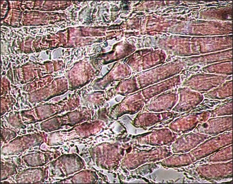
immunohistochemistry (formalin-fixed, paraffin-embedded sections): 10-20 μg/mL using porcine tongue
microarray: suitable
western blot: 0.5-1.0 μg/mL using total extract of rabbit skeletal muscle

isotype

IgG1

UniProt accession no.

shipped in

dry ice

storage temp.

−20°C

target post-translational modification

unmodified

Gene Information

Looking for similar products? Visit Product Comparison Guide

General description

Localizes an epitope on the myosin heavy chain. Stains the fast (type II) and neonatal isomyosin molecules found in skeletal muscle, but does not stain cardiac muscle, smooth muscle or non-muscle myosin in cultured cells. Does react with human rhabdomyosarcomas.

Immunogen

rabbit muscle myosin.

Application

Applications in which this antibody has been used successfully, and the associated peer-reviewed papers, are given below.
Immunohistochemistry (1 paper)
The level of mysosin (fast) in serum samples from sportsmen with past injury was determined by western blot using monoclonal mouse anti-myosin (skeletal/fast) as the primary antibody at a dilution of 1:90000.

Physical form

Solution in 0.01 M phosphate buffered saline, pH 7.4, containing 15 mM sodium azide.

Disclaimer

Unless otherwise stated in our catalog or other company documentation accompanying the product(s), our products are intended for research use only and are not to be used for any other purpose, which includes but is not limited to, unauthorized commercial uses, in vitro diagnostic uses, ex vivo or in vivo therapeutic uses or any type of consumption or application to humans or animals.

Not finding the right product?  

Try our Product Selector Tool.

Storage Class Code

10 - Combustible liquids

WGK

WGK 1


Certificates of Analysis (COA)

Search for Certificates of Analysis (COA) by entering the products Lot/Batch Number. Lot and Batch Numbers can be found on a product’s label following the words ‘Lot’ or ‘Batch’.

Already Own This Product?

Find documentation for the products that you have recently purchased in the Document Library.

Visit the Document Library

Yanlin Wang et al.
Neurological sciences : official journal of the Italian Neurological Society and of the Italian Society of Clinical Neurophysiology, 40(6), 1255-1265 (2019-03-21)
Myotonic dystrophy type 1 (DM1) is caused by CTG nucleotide repeat expansions in the 3'-untranslated region (3'-UTR) of the dystrophia myotonica protein kinase (DMPK) gene. The expanded CTG repeats encode toxic CUG RNAs that cause disease, largely through RNA gain-of-function.
Charlotte Capitanchik et al.
Nucleus (Austin, Tex.), 9(1), 410-430 (2018-06-19)
Laminopathies yield tissue-specific pathologies, yet arise from mutation of ubiquitously-expressed genes. A little investigated hypothesis to explain this is that the mutated proteins or their partners have tissue-specific splice variants. To test this, we analyzed RNA-Seq datasets, finding novel isoforms
Alan P Tenney et al.
Cell reports, 29(2), 437-452 (2019-10-10)
The somatotopic motor-neuron projections onto their cognate target muscles are essential for coordinated movement, but how that occurs for facial motor circuits, which have critical roles in respiratory and interactive behaviors, is poorly understood. We report extensive molecular heterogeneity in
Danielle Buck et al.
The Journal of general physiology, 143(2), 215-230 (2014-01-29)
Titin is a molecular spring that determines the passive stiffness of muscle cells. Changes in titin's stiffness occur in various myopathies, but whether these are a cause or an effect of the disease is unknown. We studied a novel mouse
Shirin Pourteymour et al.
Physiological reports, 3(8), doi:10-doi:10 (2015-08-13)
Perilipins (PLINs) coat the surface of lipid droplets and are important for the regulation of lipid turnover. Knowledge about the physiological role of the individual PLINs in skeletal muscle is limited although lipid metabolism is very important for muscle contraction.

Our team of scientists has experience in all areas of research including Life Science, Material Science, Chemical Synthesis, Chromatography, Analytical and many others.

Contact Technical Service