Skip to Content
Merck
All Photos(1)

Key Documents

F7884

Sigma-Aldrich

Anti-Dog IgG (whole molecule)–FITC antibody produced in rabbit

affinity isolated antibody, buffered aqueous solution

Synonym(s):

Rabbit Anti-Dog IgG (whole molecule)–Fluorescein isothiocyanate

Sign Into View Organizational & Contract Pricing


About This Item

MDL number:
UNSPSC Code:
12352203
NACRES:
NA.46

biological source

rabbit

conjugate

FITC conjugate

antibody form

affinity isolated antibody

antibody product type

secondary antibodies

clone

polyclonal

form

buffered aqueous solution

storage condition

protect from light

technique(s)

direct immunofluorescence: 1:32

storage temp.

−20°C

target post-translational modification

unmodified

Looking for similar products? Visit Product Comparison Guide

General description

IgG (Immunoglobulin G) antibody subtype is the most abundant serum immunoglobulins of the immune system. It is secreted by B cells and is found in blood and extracellular fluids and provides protection from infections caused by bacteria, fungi and viruses. Rabbit anti-dog IgG is isolated by affinity isolation and conjugated to Fluorescein Isothiocyanate (FITC).
The variable region of IgG antibody is specific to antigens and is highly conserved. IgG has four subclasses- IgG1, IgG2a, IgG2b and IgG2c.

Specificity

Anti-Dog IgG (whole molecule)-FITC antibody is specific for dog IgG subclasses.

Immunogen

Purified dog IgG

Application

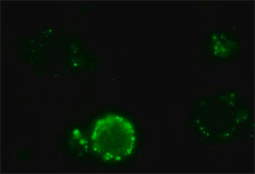
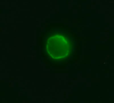
Anti-Dog IgG (whole molecule)-FITC antibody may be used for direct immunofluorescence using dog peripheral blood lymphocytes at a minimum working dilution of 1:32.

Biochem/physiol Actions

IgG (Immunoglobulin G) antibody provides protection from infections caused by bacteria, fungi and viruses. Maternal IgG is transferred to fetus through the placenta that is vital for immune defense of the neonate against infections. IgG antibody has its function similar to IgM antibody in complement system activation. IgG participates in hypersensitivity type II and type III. It helps in opsonization, complement fixation and antibody dependent cell mediated cytotoxicity.

Physical form

Solution in 0.01 M phosphate buffered saline, pH 7.4, containing 1% bovine serum albumin and 15 mM sodium azide.

Storage and Stability

For continuous use, store at 2-8 °C for up to one month.
For extended storage, the solution may be frozen in working aliquots. Repeated freezing and thawing, or storage in "frost-free" freezers, is not recommended. If slight turbidity occurs upon prolonged storage, clarify the solution by centrifugation before use.

Disclaimer

Unless otherwise stated in our catalog or other company documentation accompanying the product(s), our products are intended for research use only and are not to be used for any other purpose, which includes but is not limited to, unauthorized commercial uses, in vitro diagnostic uses, ex vivo or in vivo therapeutic uses or any type of consumption or application to humans or animals.

Not finding the right product?  

Try our Product Selector Tool.

Storage Class Code

10 - Combustible liquids

WGK

nwg

Flash Point(F)

Not applicable

Flash Point(C)

Not applicable

Personal Protective Equipment

dust mask type N95 (US), Eyeshields, Gloves

Certificates of Analysis (COA)

Search for Certificates of Analysis (COA) by entering the products Lot/Batch Number. Lot and Batch Numbers can be found on a product’s label following the words ‘Lot’ or ‘Batch’.

Already Own This Product?

Find documentation for the products that you have recently purchased in the Document Library.

Visit the Document Library

Vanessa Figueredo Pereira et al.
Revista da Sociedade Brasileira de Medicina Tropical, 49(1), 104-106 (2016-05-11)
Conjunctival swab PCR was evaluated as a tool to diagnose visceral leishmaniasis in dogs. Conjunctival swab PCR was compared to indirect immunofluorescence antibody test and blood PCR. Indirect immunofluorescence was significantly correlated with conjunctival swab PCR (p < 0.05), but
Hanna D Bremer et al.
Scientific reports, 8(1), 4852-4852 (2018-03-21)
Dogs can spontaneously develop complex systemic autoimmune disorders, with similarities to human autoimmune disease. Autoantibodies directed at self-antigens are a key feature of these autoimmune diseases. Here we report the identification of interleukin enhancer-binding factors 2 and 3 (ILF2 and
Gisele Vaz Aguirre Samoel et al.
Revista brasileira de parasitologia veterinaria = Brazilian journal of veterinary parasitology : Orgao Oficial do Colegio Brasileiro de Parasitologia Veterinaria, 33(1), e013723-e013723 (2023-12-21)
Bovine trypanosomosis, caused by Trypanosoma vivax, is a disease that originated in Africa and currently affects cattle in several South American countries, including almost all Brazilian states. Despite the reports on T. vivax infection in southern Brazil, data on its
Comparative evaluation of the DPP? CVL rapid test for canine serodiagnosis in area of visceral leishmaniasis.
Laurenti M D, et al.
Veterinary Parasitology, 205(3-4) (2014)
Nataša Knap et al.
Viruses, 12(7) (2020-07-09)
West Nile virus (WNV) is a flavivirus transmitted by mosquitoes. Birds are the reservoir for the virus; humans, horses and other mammals are dead-end hosts. Infections caused by WNV in humans can vary from asymptomatic infections to West Nile fever

Our team of scientists has experience in all areas of research including Life Science, Material Science, Chemical Synthesis, Chromatography, Analytical and many others.

Contact Technical Service