Skip to Content
Merck
All Photos(2)

Key Documents

A5691

Sigma-Aldrich

Anti-Actin, α-Smooth Muscle - Alkaline Phosphatase antibody, Mouse monoclonal

clone 1A4, purified from hybridoma cell culture

Synonym(s):

Monoclonal Anti-Actin, α-Smooth Muscle, Monoclonal Anti-Actin, α-Smooth Muscle - Alkaline Phosphatase antibody produced in mouse, SMA

Sign Into View Organizational & Contract Pricing


About This Item

UNSPSC Code:
12352203
NACRES:
NA.41

biological source

mouse

Quality Level

conjugate

alkaline phosphatase conjugate

antibody form

purified from hybridoma cell culture

antibody product type

primary antibodies

clone

1A4, monoclonal

form

buffered aqueous glycerol solution

mol wt

antigen ~42 kDa

species reactivity

human, mouse, rat, chicken, frog, canine, rabbit, guinea pig, goat, bovine, sheep, snake

technique(s)

ELISA: suitable
immunohistochemistry (formalin-fixed, paraffin-embedded sections): 1:20 using human tonsil or appendix sections
western blot: 1:100 using chicken gizzard extract/ Mouse heart extract

isotype

IgG2a

UniProt accession no.

shipped in

wet ice

storage temp.

2-8°C

target post-translational modification

unmodified

Gene Information

mouse ... Acta2(11475)
rat ... Acta2(81633)

Looking for similar products? Visit Product Comparison Guide

General description

Actin is a highly conserved protein that is a major component of both the cytoskeletal and contractile structures in all cell types. It varies in amount, being related to the type of differentiation and to the functional state of cells and tissues. Actin can be found in two different forms of aggregation, the globular or the fibrillar state, and at least six distinct isoforms occur in vertebrates. The actins exhibit over 90% sequence homology, but each isoform has a unique NH2-terminal sequence. The isoforms are comprised of three α actins (skeletal, cardiac, smooth), one β actin (β-non-muscle) and two γ actins (γ smooth muscle and γ non-muscle).
The antibody (also known as anti-α-Sm-1) is specific for the single isoform of α-smooth muscle actin. It reacts specifically with α-smooth muscle actin in immunoblotting assays and labels smooth muscle cells in frozen or formalin-fixed, paraffin-embedded tissue sections.

Immunogen

N-terminal synthetic decapeptide of α-smooth muscle actin.

Application

Applications in which this antibody has been used successfully, and the associated peer-reviewed papers, are given below.
Immunohistochemistry (1 paper)
Immunocytochemistry was performed on kertocytes fixed in 1% PFA and incubated with mouse monoclonal anti-smooth muscle actin (14A) at a 1:100 dilution.
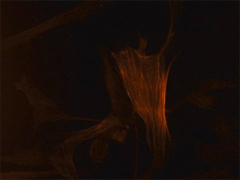
Immunocytochemistry was performed on smooth muscle cells from bovine aortas using the monoclonal anti-ACTA2 antibody. Cells were first grown on glass cover slips and fixed in 50% acetone/EtOH for 10 minutes at 4 degrees.
Mouse monoclonal anti-actin, α−smooth muscle-alkaline phosphatase antibody has been used for immunohistochemical application in mouse and human tissues.
Paraffin embedded sections of rat testis tissue grafts were immunohistochemically stained with mouse monoclonal anti-smooth muscle actin.
IHC analysis of x-gal stained muouse cardiac tissue was performed using the primary antibody, mouse monoclonal anti-smooth muscle actin to identify myofibroblasts.

Physical form

Solution in 0.05 M Tris buffer, pH 8.0, containing 1 mM MgCl2, 1% bovine serum albumin, 50% glycerol, and 15 mM sodium azide as a preservative.

Other Notes

To view an Actin antibody selection guide, please visit www.sigmaaldrich.com/actin.

Disclaimer

Unless otherwise stated in our catalog or other company documentation accompanying the product(s), our products are intended for research use only and are not to be used for any other purpose, which includes but is not limited to, unauthorized commercial uses, in vitro diagnostic uses, ex vivo or in vivo therapeutic uses or any type of consumption or application to humans or animals.

Not finding the right product?  

Try our Product Selector Tool.

Storage Class Code

10 - Combustible liquids

WGK

WGK 2

Personal Protective Equipment

dust mask type N95 (US), Eyeshields, Gloves

Certificates of Analysis (COA)

Search for Certificates of Analysis (COA) by entering the products Lot/Batch Number. Lot and Batch Numbers can be found on a product’s label following the words ‘Lot’ or ‘Batch’.

Already Own This Product?

Find documentation for the products that you have recently purchased in the Document Library.

Visit the Document Library

Maryellen Sandor et al.
Eplasty, 17, e1-e1 (2017-01-26)
Objective: Benchtop methods were evaluated for preclinical inflammation/capsule formation correlation following implantation of human acellular dermal matrices. Methods: Dermal matrices were compared with native dermis for structure (histology, scanning electron microscopy), collagen solubility (hydroxyproline), enzymatic susceptibility (collagenase), and thermal stability
J V Jester et al.
Investigative ophthalmology & visual science, 40(9), 1959-1967 (1999-08-10)
Recent studies indicate that transforming growth factor (TGF)beta is a potent inducer of corneal myofibroblast differentiation and expression of smooth muscle-specific, alpha-actin (alpha-SMA). Although TGFbeta is known to enhance synthesis of extracellular matrix proteins and receptors, little is known about
Kenneth Chan et al.
Journal of the American Heart Association, 6(11) (2017-11-02)
Genome-wide association studies identified ADAMTS7 as a risk locus for coronary artery disease (CAD). Functional studies suggest that ADAMTS7 may promote cellular processes in atherosclerosis. We sought to examine the association between genetic variation at ADAMTS7 and measures of atherosclerosis
Xiaoqing Guo et al.
The American journal of pathology, 179(3), 1385-1393 (2011-07-19)
Ovarian carcinoma is the most lethal gynecologic malignancy, however underlying molecular events remain elusive. Expression of human chorionic gonadotropin β subunit (β-hCG) is clinically significant for both trophoblastic and nontrophoblastic cancers; however, whether β-hCG facilitates ovarian epithelial cell tumorigenic potential
Alex H P Chan et al.
PloS one, 12(3), e0174773-e0174773 (2017-03-30)
Current animal models for the evaluation of synthetic grafts are lacking many of the molecular tools and transgenic studies available to other branches of biology. A mouse model of vascular grafting would allow for the study of molecular mechanisms of

Our team of scientists has experience in all areas of research including Life Science, Material Science, Chemical Synthesis, Chromatography, Analytical and many others.

Contact Technical Service