Skip to Content
Merck
All Photos(1)

Key Documents

MABN1797

Sigma-Aldrich

Anti-Prdx-6 Antibody, clone TAG-2A12

clone TAG-2A12, from mouse

Synonym(s):

Peroxiredoxin-6, EC: 1.11.1.15, 1-Cys peroxiredoxin, 1-Cys PRX, 24 kDa protein, Acidic calcium-independent phospholipase A2, aiPLA2, Antioxidant protein 2, Liver 2D page spot 40, Non-selenium glutathione peroxidase, NSGPx

Sign Into View Organizational & Contract Pricing


About This Item

UNSPSC Code:
12352203
eCl@ss:
32160702
NACRES:
NA.41
clone:
TAG-2A12, monoclonal
application:
FACS
ICC
IF
IP
WB
species reactivity:
human
technique(s):
flow cytometry: suitable
immunocytochemistry: suitable
immunofluorescence: suitable
immunoprecipitation (IP): suitable
western blot: suitable
citations:
2

biological source

mouse

Quality Level

antibody form

purified immunoglobulin

antibody product type

primary antibodies

clone

TAG-2A12, monoclonal

species reactivity

human

packaging

antibody small pack of 25 μg

technique(s)

flow cytometry: suitable
immunocytochemistry: suitable
immunofluorescence: suitable
immunoprecipitation (IP): suitable
western blot: suitable

isotype

IgG1κ

NCBI accession no.

UniProt accession no.

shipped in

ambient

target post-translational modification

unmodified

Gene Information

human ... PRDX6(9588)

General description

Peroxiredoxin-6 (UniProt: P30041; also known as EC: 1.11.1.15, 1-Cys peroxiredoxin, 1-Cys PRX, 24 kDa protein, Acidic calcium-independent phospholipase A2, aiPLA2, Antioxidant protein 2, Liver 2D page spot 40, Non-selenium glutathione peroxidase, NSGPx) is encoded by the PRDX6 (also known as AOP2, KIAA0106) gene (Gene ID: 9588) in human. Peroxiredoxin-6 is a cytoplasmic protein that is involved in redox regulation of the cell. It reduces hydrogen peroxide and short chain organic fatty acid, and phospholipid hydroperoxides. It is the only mammalian 1-Cys member of the Prdx family with bifunctional - glutathione peroxidase and phospholipase A2 activities. Peroxiredoxin-6 shares several structural and functional properties with other members of the Prdx family, however, it has only one conserved cysteine residue and its thioredoxin does not participate in the catalytic cycle. Peroxiredoxin-6 is expressed in higher levels in epithelium such as apical regions of respiratory epithelium and skin epidermis and within the lung, it is expressed at relatively high levels within the type II alveolar epithelial cells and bronchiolar Clara cells. Lower levels of expression are reported in alveolar macrophages and microvascular endothelium. Human peroxiredoxin-6 shares 87% sequence identity with mouse, rat, and bovine.

Specificity

Clone TAG-2A12 detects Peroxiredoxin-6 in human cells.

Immunogen

Corneal endothelial cells (hCENC) isolated from corneas of human cadavers and immunized with monophosphoryl-lipid A trehalose dicorynomycolate (MPLCTDM) adjuvant.

Application

Anti-Prdx-6 Antibody, clone TAG-2A12, Cat. No. MABN1797, is a highly specific mouse monoclonal antibody that targets Prdx-6 and has been tested in Flow Cytometry, Immunofluorescence, Immunocytochemistry and Immunoprecipitation, and Western Blotting.
Immunoprecipitation Analysis: A representative lot detected Prdx-6 in Immunoprecipitation applications (Ding, V., et al. (2014). MAbs. 6(6):1439-52).

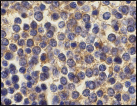
Immunocytochemistry Analysis: A representative lot detected Prdx-6 in Immunocytochemistry applications (Ding, V., et al. (2014). MAbs. 6(6):1439-52).

Flow Cytometry Analysis: A representative lot detected Prdx-6 in Flow Cytometry applications (Ding, V., et al. (2014). MAbs. 6(6):1439-52).

Immunofluorescence Analysis: A representative lot detected Prdx-6 in Immunofluorescence applications (Ding, V., et al. (2014). MAbs. 6(6):1439-52).

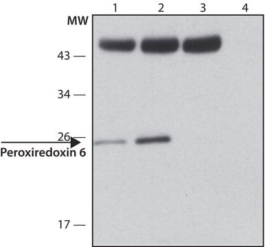
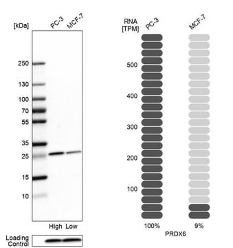
Western Blotting Analysis: A representative lot detected Prdx-6 in Western Blotting applications (Ding, V., et al. (2014). MAbs. 6(6):1439-52).

Flow Cytometry Analysis: A representative lot detected Prdx-6 in human corneal endothelial cells from a donor. (Courtesy of Dr. Vanessa Ding, Ph.D. and Dr. Andre Choo, Ph.D, BTI, Singapore).
Research Category
Neuroscience

Quality

Identity Confirmation by Isotyping Test.

Isotyping Analysis: The identity of this monoclonal antibody is confirmed by isotyping test to be human IgG1 .

Target description

~28 kDa observed; 25.03 kDa calculated. Uncharacterized bands may be observed in some lysate(s).

Physical form

Format: Purified
Protein G purified
Purified mouse monoclonal antibody IgG1 in buffer containing 0.1 M Tris-Glycine (pH 7.4), 150 mM NaCl with 0.05% sodium azide.

Storage and Stability

Stable for 1 year at 2-8°C from date of receipt.

Other Notes

Concentration: Please refer to lot specific datasheet.

Disclaimer

Unless otherwise stated in our catalog or other company documentation accompanying the product(s), our products are intended for research use only and are not to be used for any other purpose, which includes but is not limited to, unauthorized commercial uses, in vitro diagnostic uses, ex vivo or in vivo therapeutic uses or any type of consumption or application to humans or animals.

Not finding the right product?  

Try our Product Selector Tool.

Storage Class Code

12 - Non Combustible Liquids

WGK

WGK 1

Flash Point(F)

Not applicable

Flash Point(C)

Not applicable


Certificates of Analysis (COA)

Search for Certificates of Analysis (COA) by entering the products Lot/Batch Number. Lot and Batch Numbers can be found on a product’s label following the words ‘Lot’ or ‘Batch’.

Already Own This Product?

Find documentation for the products that you have recently purchased in the Document Library.

Visit the Document Library

Julia María Torres-Velarde et al.
Free radical biology & medicine, 218, 82-93 (2024-04-06)
Peroxiredoxin 6 (Prdx6) repairs peroxidized membranes by reducing oxidized phospholipids, and by replacing oxidized sn-2 fatty acyl groups through hydrolysis/reacylation by its phospholipase A2 (aiPLA2) and lysophosphatidylcholine acyltransferase activities. Prdx6 is highly expressed in the lung, and intact lungs and
Sylwia Bobis-Wozowicz et al.
Cell communication and signaling : CCS, 22(1), 356-356 (2024-07-10)
Stem cell-derived extracellular vesicles (EVs) are an emerging class of therapeutics with excellent biocompatibility, bioactivity and pro-regenerative capacity. One of the potential targets for EV-based medicines are cardiovascular diseases (CVD). In this work we used EVs derived from human induced

Our team of scientists has experience in all areas of research including Life Science, Material Science, Chemical Synthesis, Chromatography, Analytical and many others.

Contact Technical Service